Department of Radiology, Altınbas University School of Medicine Bahcelievler Medical Park Hospital, İstanbul, Turkey.
Department of Radiology, University of Health Sciences, Prof Dr Cemil Tascıoglu City Hospital, Istanbul, Turkey.
BMC Pediatr. 2021 Oct 11;21(1):445. doi: 10.1186/s12887-021-02890-y.
Haematopoietic stem cell transplantation (HSCT) is used worldwide in various malignant and nonmalignant childhood diseases, including haematologic, genetic, autoimmune and metabolic disorders, and is the only curative treatment for many of these illnesses. The survival rates of many childhood diseases have been increased due to HSCT treatment. However, associated complications are still important for management. Central nervous system (CNS) complications in paediatric HSCT recipients can be associated with high morbidity and significantly contribute to mortality. Posterior reversible encephalopathy syndrome (PRES) is one of the most common CNS complications in patients with neurological symptoms following HSCT. Magnetic resonance imaging (MRI) is the modality of choice and shows typical bilateral vasogenic oedema at the posterior parts of the cerebral hemispheres; however, various atypical imaging manifestations can also occur. In this study, we retrospectively examined CNS complications in our paediatric HSCT recipients with a focus on the typical and atypical neuroimaging manifestations of PRES following HSCT.
We retrospectively reviewed the medical records of 300 consecutive paediatric HSCT recipients from January 2014 to November 2018. A total of 130 paediatric HSCT recipients who experienced neurological signs and symptoms and were evaluated with neuroimaging studies following HSCT were enrolled in the study. The timing of CNS complications was defined according to immune status, including the pre-engraftment period (< 30 days after HSCT), the early postengraftment period (30-100 days after HSCT), and the late postengraftment period (> 100 days after HSCT), which were defined as phases 1, 2 and 3, respectively.
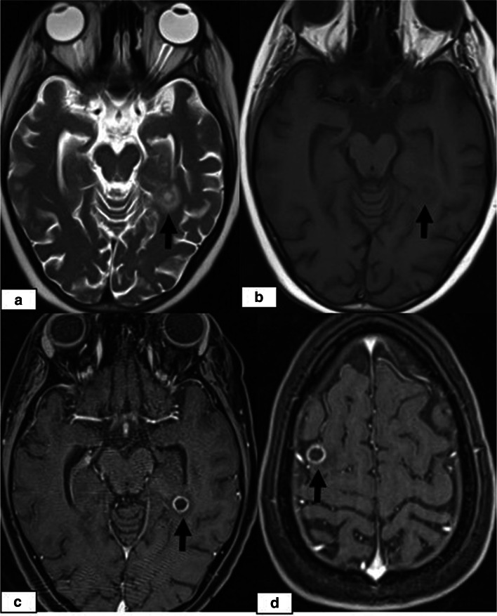
Overall, 130 paediatric HSCT recipients experienced neurological signs and symptoms and therefore underwent neuroimaging examinations. Among these 130 patients, CNS complications were present in 23 patients (17.6%, 23/130), including 13 (56.5%) females and 10 (43.5%) males with a median age of 8.0 years (range, 8 months to 18.0 years). Among these 23 patients, 14 cases of PRES (60.9%), 5 (21.7%) cases of leukoencephalopathy, 3 cases of acute subdural haemorrhage (ASDH) (13%) and 1 (4.3%) case of fungal CNS infection were identified by neuroimaging. On MRI, typical parietooccipital vasogenic oedema was present in 78.5% of the PRES cases (11/14). The following atypical neuroimaging manifestations were observed: isolated involvement of the bilateral frontal lobes in 1 case, isolated cerebellar vermis involvement in 1 case, and isolated basal ganglia involvement in 1 case. Restricted diffusion associated with cytotoxic damage was demonstrated in 2 of 14 cases, one of which also showed subacute cytotoxic injury with ADC pseudonormalization.
Paediatric HSCT recipients presenting with CNS signs and symptoms should be evaluated by neuroimaging studies for timely diagnosis and early management. PRES is the most common CNS complication and may present with atypical MRI manifestations, which should not dissuade a PRES diagnosis in appropriate clinical settings.

造血干细胞移植(HSCT)在全球范围内用于治疗各种儿童期恶性和非恶性疾病,包括血液、遗传、自身免疫和代谢疾病,是许多此类疾病的唯一治愈方法。由于 HSCT 治疗,许多儿童疾病的存活率已经提高。然而,相关并发症仍然是管理的重点。儿科 HSCT 受者的中枢神经系统(CNS)并发症可能与高发病率有关,并显著导致死亡率。移植后脑病综合征(PRES)是 HSCT 后出现神经症状的患者中最常见的 CNS 并发症之一。磁共振成像(MRI)是首选的方法,显示典型的双侧血管源性水肿位于大脑半球后部;然而,也可能出现各种非典型的影像学表现。在这项研究中,我们回顾性检查了我们儿科 HSCT 受者的 CNS 并发症,重点关注 HSCT 后 PRES 的典型和非典型神经影像学表现。
我们回顾性分析了 2014 年 1 月至 2018 年 11 月期间连续 300 例儿科 HSCT 受者的病历。共纳入 130 例出现神经症状并在 HSCT 后接受神经影像学检查的儿科 HSCT 受者。根据免疫状态定义 CNS 并发症的时间,包括植入前期(HSCT 后<30 天)、早期植入后期(HSCT 后 30-100 天)和晚期植入后期(HSCT 后>100 天),分别定义为第 1、2 和 3 期。
共有 130 例儿科 HSCT 受者出现神经症状,因此进行了神经影像学检查。在这 130 例患者中,23 例(17.6%,23/130)出现 CNS 并发症,包括 13 例(56.5%)女性和 10 例(43.5%)男性,中位年龄为 8.0 岁(范围为 8 个月至 18.0 岁)。在这 23 例患者中,通过神经影像学检查发现 PRES 14 例(60.9%)、白质脑病 5 例(21.7%)、急性硬膜下血肿(ASDH)3 例(13%)和真菌性 CNS 感染 1 例(4.3%)。在 MRI 上,78.5%(11/14)的 PRES 病例存在典型的顶枕叶血管源性水肿。观察到以下非典型神经影像学表现:1 例双侧额叶孤立受累,1 例小脑蚓部孤立受累,1 例基底节孤立受累。在 14 例中,2 例显示细胞毒性损伤相关的弥散受限,其中 1 例也表现为亚急性细胞毒性损伤,ADC 假性正常化。
出现 CNS 症状的儿科 HSCT 受者应通过神经影像学检查进行评估,以便及时诊断和早期治疗。PRES 是最常见的 CNS 并发症,可能表现为非典型 MRI 表现,但在适当的临床环境下不应排除 PRES 诊断。