Neuroscience Graduate Program, University of Rochester Medical Center, 601 Elmwood Ave. Box 603, Rochester, NY, 14642, USA.
Center for Neural Science, New York University, New York, NY, 10003, USA.
Brain Struct Funct. 2021 Dec;226(9):2777-2791. doi: 10.1007/s00429-021-02385-7. Epub 2021 Oct 12.
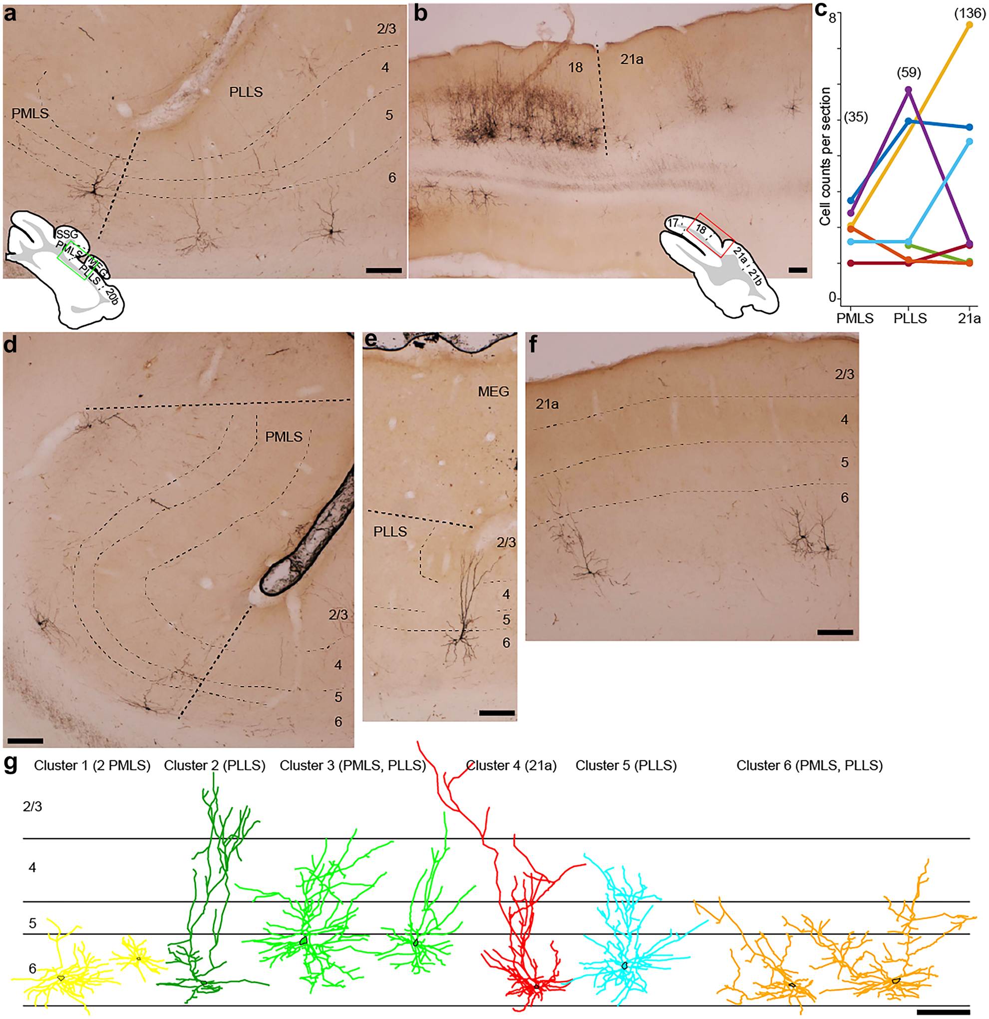
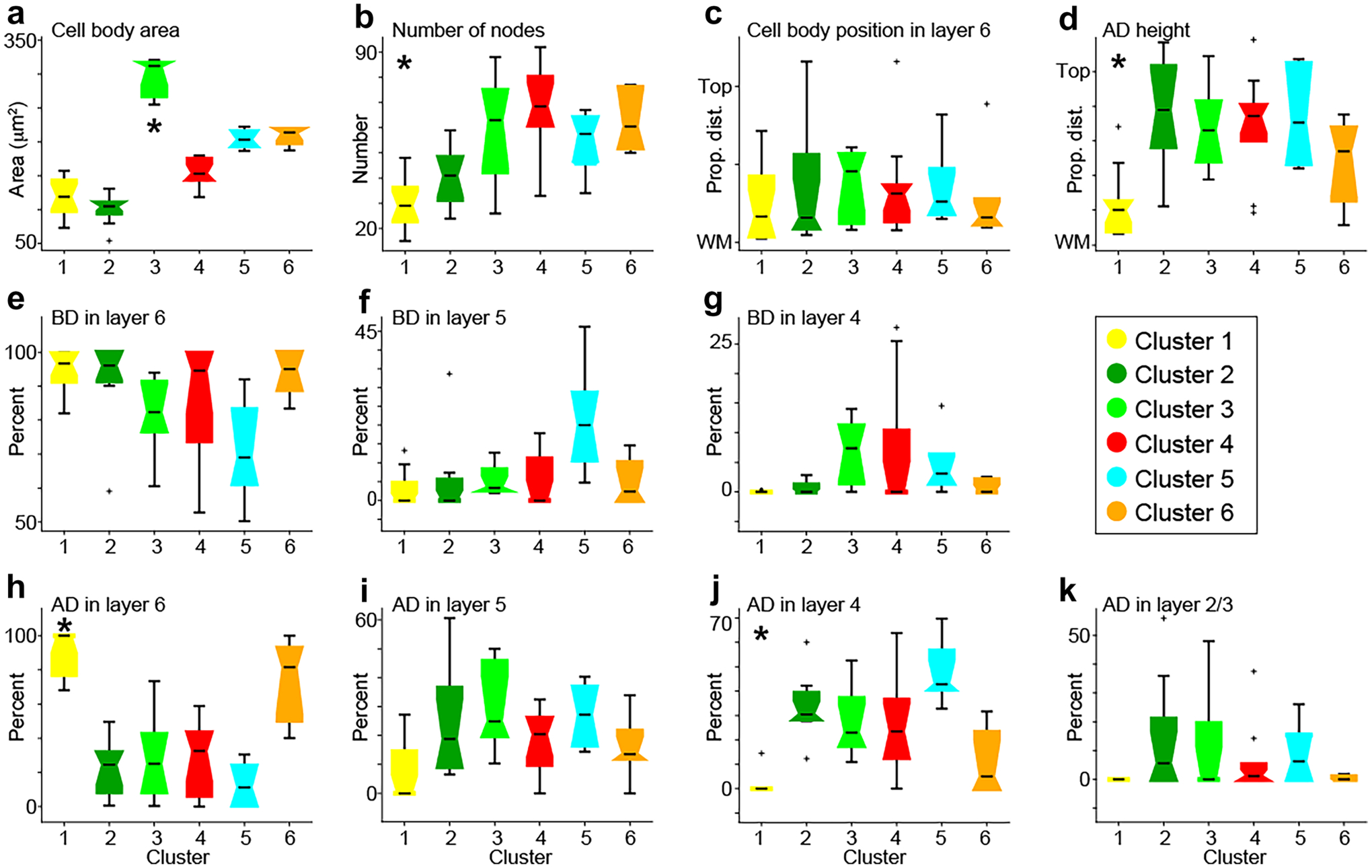
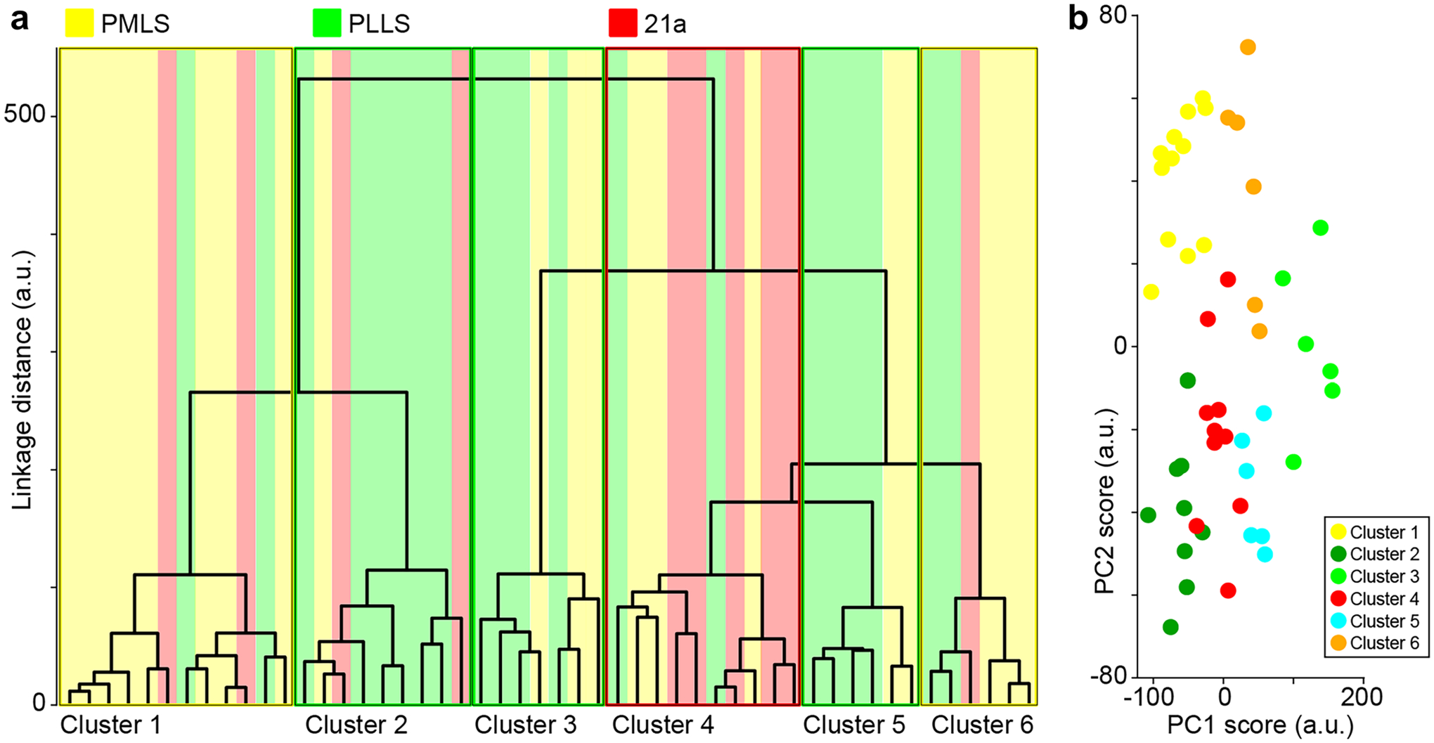
Complementary reciprocal feedforward and feedback circuits connecting the visual thalamus with the visual cortex are essential for visual perception. These circuits predominantly connect primary and secondary visual cortex with the dorsal lateral geniculate nucleus (LGN). Although there are direct geniculocortical inputs to extrastriate visual cortex, whether reciprocal corticogeniculate neurons exist in extrastriate cortex is not known. Here we utilized virus-mediated retrograde tracing to reveal the presence of corticogeniculate neurons in three mid-level extrastriate visual cortical areas in ferrets: PMLS, PLLS, and 21a. We observed corticogeniculate neurons in all three extrastriate areas, although the density of virus-labeled corticogeniculate neurons in extrastriate cortex was an order of magnitude less than that in areas 17 and 18. A cluster analysis of morphological metrics quantified following reconstructions of the full dendritic arborizations of virus-labeled corticogeniculate neurons revealed six distinct cell types. Similar corticogeniculate cell types to those observed in areas 17 and 18 were also observed in PMLS, PLLS, and 21a. However, these unique cell types were not equally distributed across the three extrastriate areas. The majority of corticogeniculate neurons per cluster originated in a single area, suggesting unique parallel organizations for corticogeniculate feedback from each extrastriate area to the LGN. Together, our findings demonstrate direct feedback connections from mid-level extrastriate visual cortex to the LGN, supporting complementary reciprocal circuits at multiple processing stages along the visual hierarchy. Importantly, direct reciprocal connections between the LGN and extrastriate cortex, that bypass V1, could provide a substrate for residual vision following V1 damage.
连接视丘脑与视皮层的互补互馈前馈和反馈回路对视知觉至关重要。这些回路主要将初级和次级视皮层与背外侧膝状体核(LGN)连接起来。尽管外纹状皮层有直接的视束传入,但外纹状皮层中是否存在回返皮质-膝状体神经元尚不清楚。在这里,我们利用病毒介导的逆行追踪来揭示雪貂三个中等级视皮层区域(PMLS、PLLS 和 21a)中存在皮质-膝状体神经元。我们在所有三个外纹状皮层区域都观察到了皮质-膝状体神经元,尽管外纹状皮层中病毒标记的皮质-膝状体神经元的密度比 17 和 18 区低一个数量级。对病毒标记的皮质-膝状体神经元全树突分支进行重建后,对形态学指标进行聚类分析,定量分析了六个不同的细胞类型。在 PMLS、PLLS 和 21a 中观察到的与 17 和 18 区观察到的相似的皮质-膝状体细胞类型,也观察到了与 17 和 18 区观察到的相似的皮质-膝状体细胞类型。然而,这些独特的细胞类型在三个外纹状区域并不均匀分布。每个簇的大多数皮质-膝状体神经元起源于单个区域,这表明从每个外纹状区域到 LGN 的皮质-膝状体反馈具有独特的平行组织。总之,我们的发现表明从中等级视皮层到 LGN 的直接反馈连接,支持视觉层次结构中多个处理阶段的互补互馈回路。重要的是,LGN 和外纹状皮层之间的直接回返连接,绕过 V1,可为 V1 损伤后的残留视觉提供一个基础。