Section on Cognitive Neurophysiology and Imaging, National Institute of Mental Health, National Institutes of Health, Bethesda, MD 20892.
Neurophysiology Imaging Facility, National Institute of Mental Health, National Institute for Neurological Disorders and Stroke, National Eye Institute, National Institutes of Health, Bethesda, MD 20892.
Proc Natl Acad Sci U S A. 2022 Sep 6;119(36):e2206559119. doi: 10.1073/pnas.2206559119. Epub 2022 Aug 31.
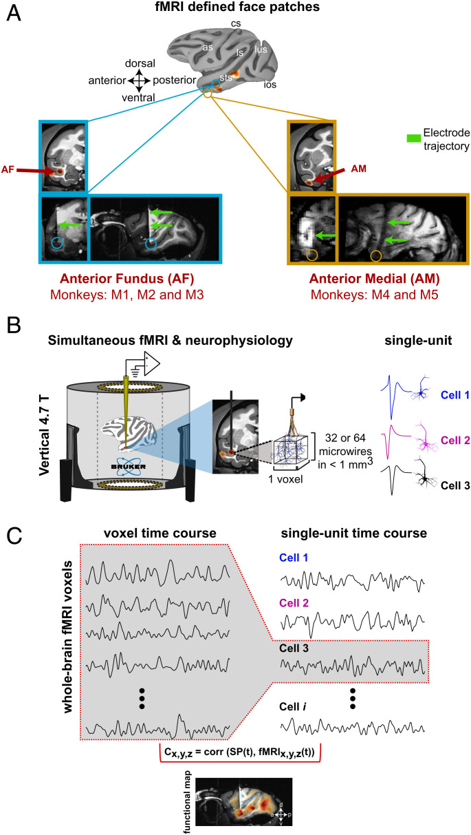
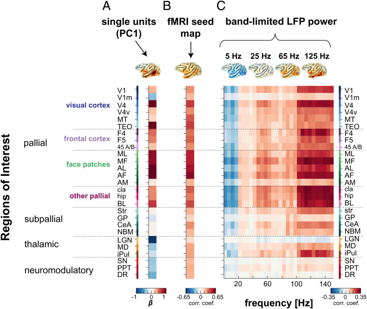
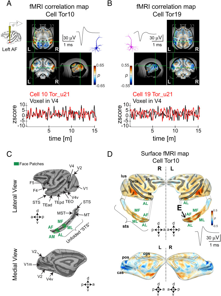
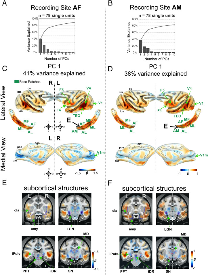
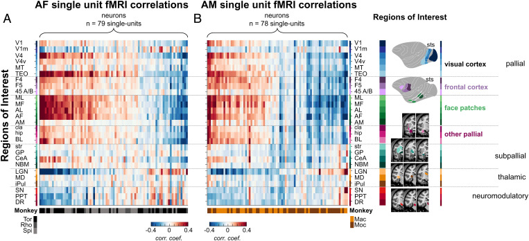
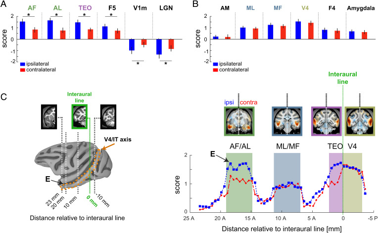
The brain is a highly organized, dynamic system whose network architecture is often assessed through resting functional magnetic resonance imaging (fMRI) functional connectivity. The functional interactions between brain areas, including those observed during rest, are assumed to stem from the collective influence of action potentials carried by long-range neural projections. However, the contribution of individual neurons to brain-wide functional connectivity has not been systematically assessed. Here we developed a method to concurrently measure and compare the spiking activity of local neurons with fMRI signals measured across the brain during rest. We recorded spontaneous activity from neural populations in cortical face patches in the macaque during fMRI scanning sessions. Individual cells exhibited prominent, bilateral coupling with fMRI fluctuations in a restricted set of cortical areas inside and outside the face patch network, partially matching the pattern of known anatomical projections. Within each face patch population, a subset of neurons was positively coupled with the face patch network and another was negatively coupled. The same cells showed inverse correlations with distinct subcortical structures, most notably the lateral geniculate nucleus and brainstem neuromodulatory centers. Corresponding connectivity maps derived from fMRI seeds and local field potentials differed from the single unit maps, particularly in subcortical areas. Together, the results demonstrate that the spiking fluctuations of neurons are selectively coupled with discrete brain regions, with the coupling governed in part by anatomical network connections and in part by indirect neuromodulatory pathways.
大脑是一个高度组织化、动态的系统,其网络结构通常通过静息功能磁共振成像(fMRI)功能连接来评估。大脑区域之间的功能相互作用,包括在休息期间观察到的相互作用,被认为源自长程神经投射携带的动作电位的集体影响。然而,个别神经元对全脑功能连接的贡献尚未得到系统评估。在这里,我们开发了一种方法,可在静息期间同时测量和比较局部神经元的尖峰活动与整个大脑的 fMRI 信号。我们在猕猴的大脑皮层面部斑块中记录了在 fMRI 扫描过程中的自发活动。单个细胞表现出与 fMRI 波动的显著、双侧耦合,这种耦合局限于面部斑块网络内部和外部的一组皮质区域,部分匹配已知解剖学投射的模式。在每个面部斑块群体中,一部分神经元与面部斑块网络呈正相关,另一部分神经元与面部斑块网络呈负相关。同一细胞与不同的皮质下结构呈相反的相关性,尤其是外侧膝状体核和脑干神经调质中心。源自 fMRI 种子和局部场电位的相应连接图与单细胞图谱不同,特别是在皮质下区域。总之,这些结果表明,神经元的尖峰波动与离散的脑区选择性耦合,这种耦合部分由解剖网络连接决定,部分由间接神经调质途径决定。