Ichida Jennifer M, Mavity-Hudson Julia A, Casagrande Vivien A
Department of Psychology, Vanderbilt University, Nashville, TN, USA.
Department of Cell and Developmental Biology, Vanderbilt University, Nashville, TN, USA.
Eye Brain. 2014 Sep;2014(6 Suppl 1):57-73. doi: 10.2147/EB.S64281.
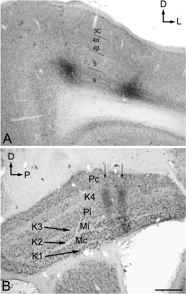
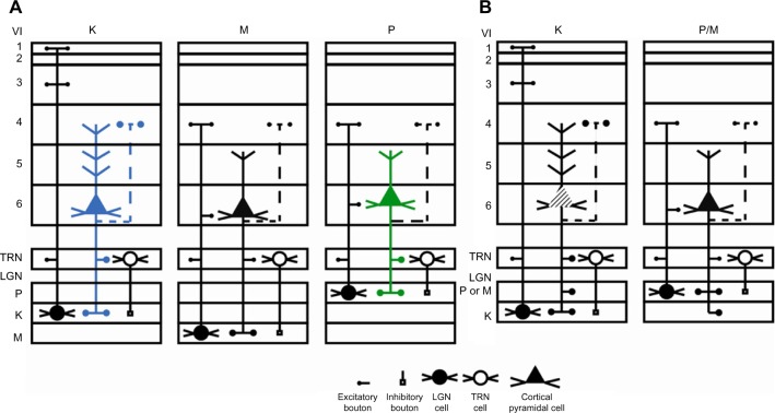
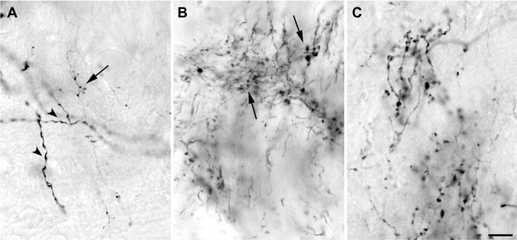
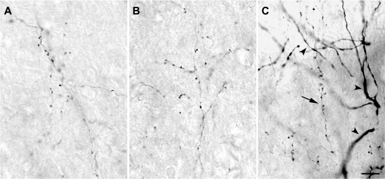
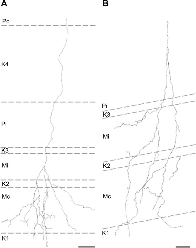
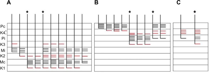
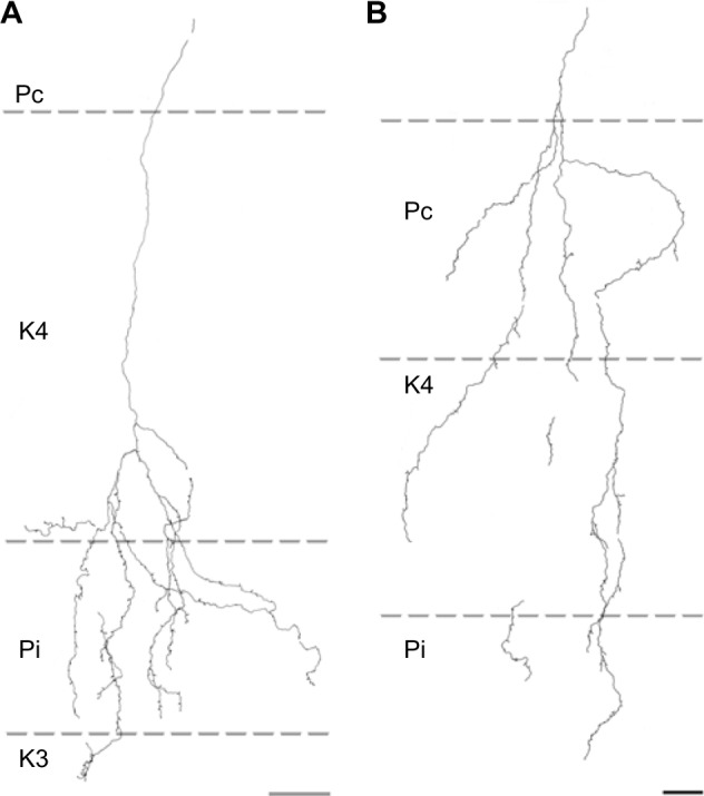
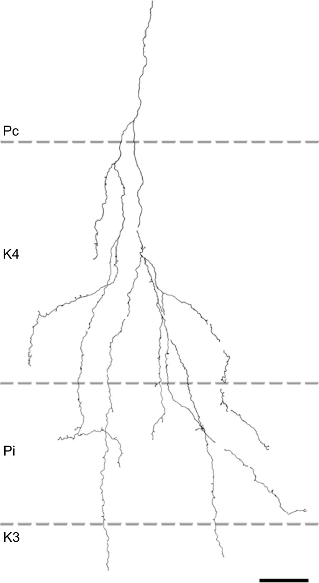
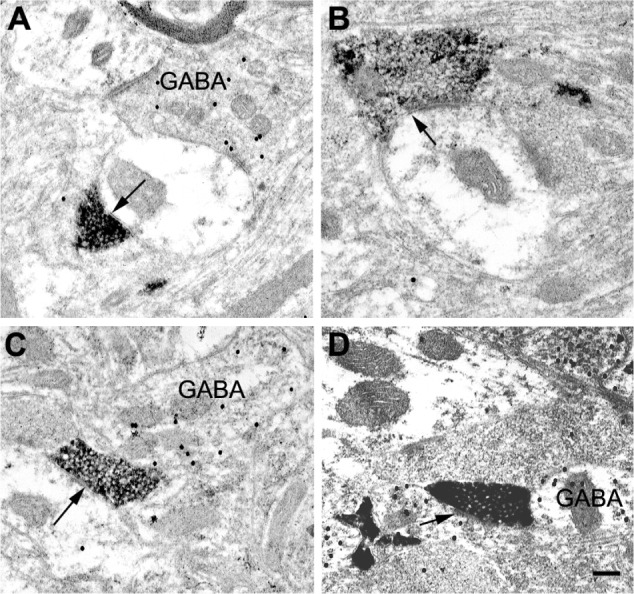
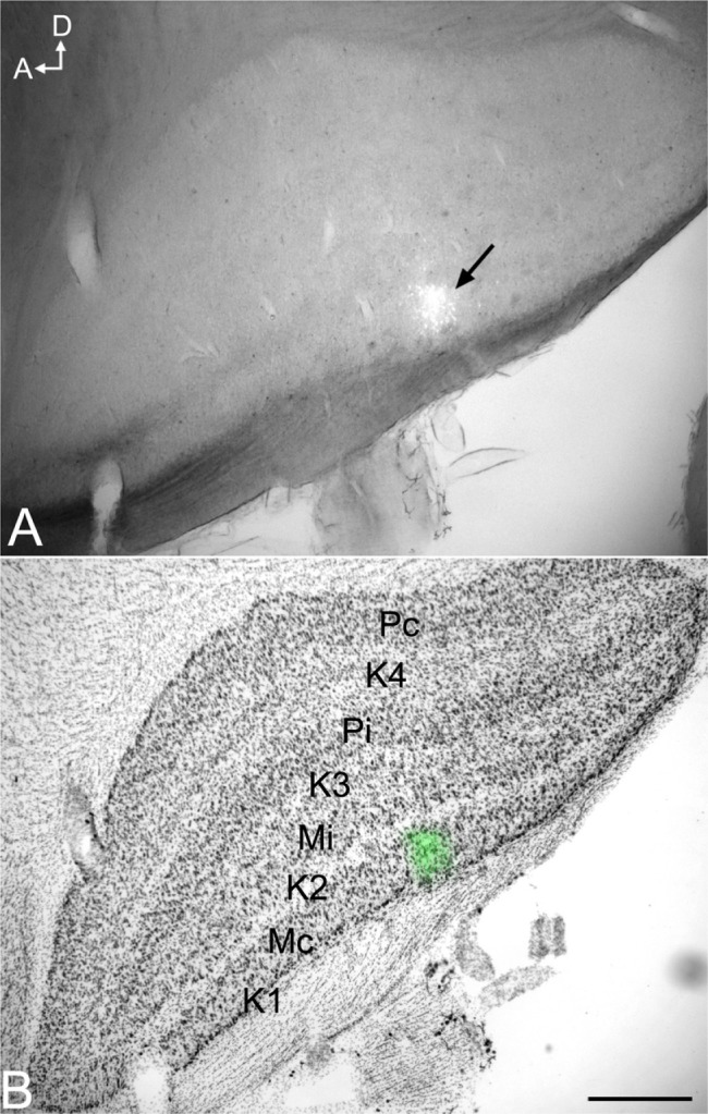
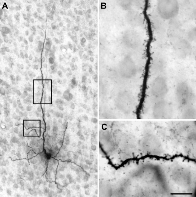
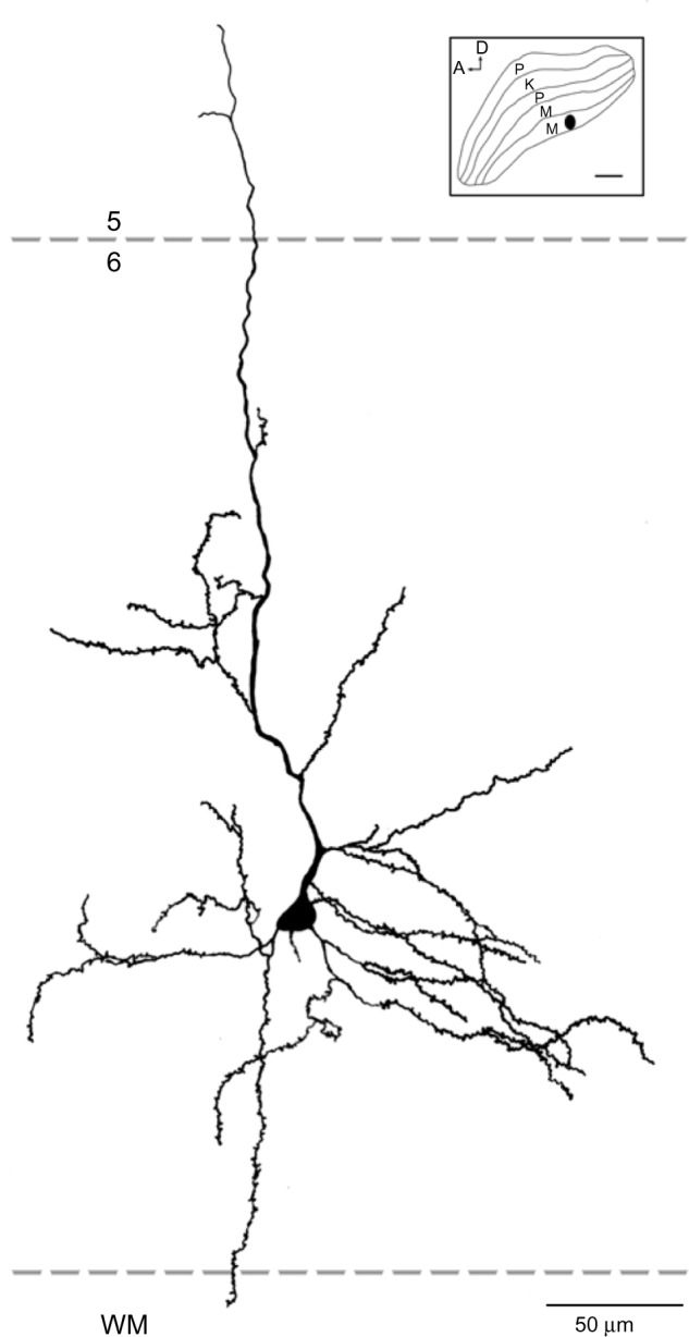
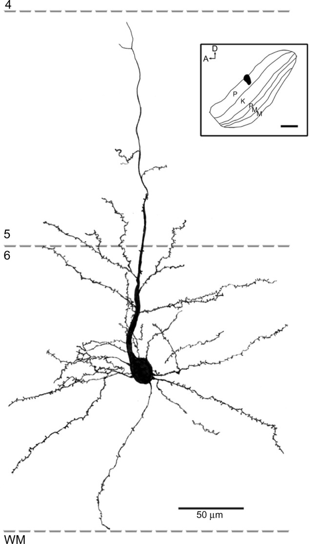
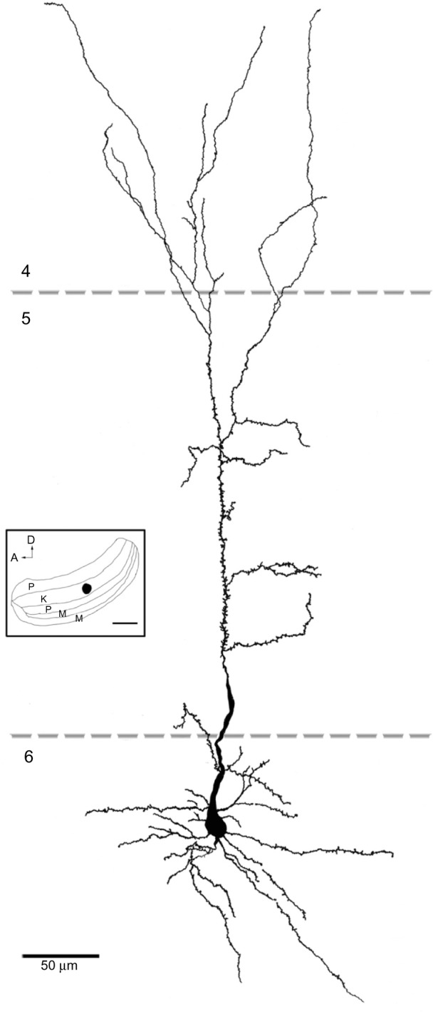
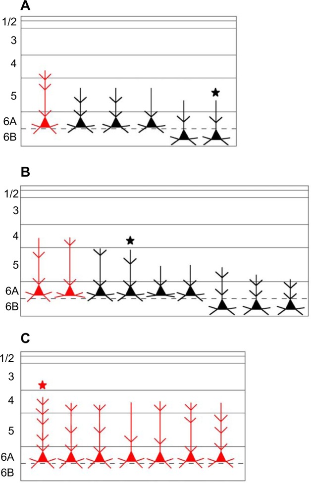
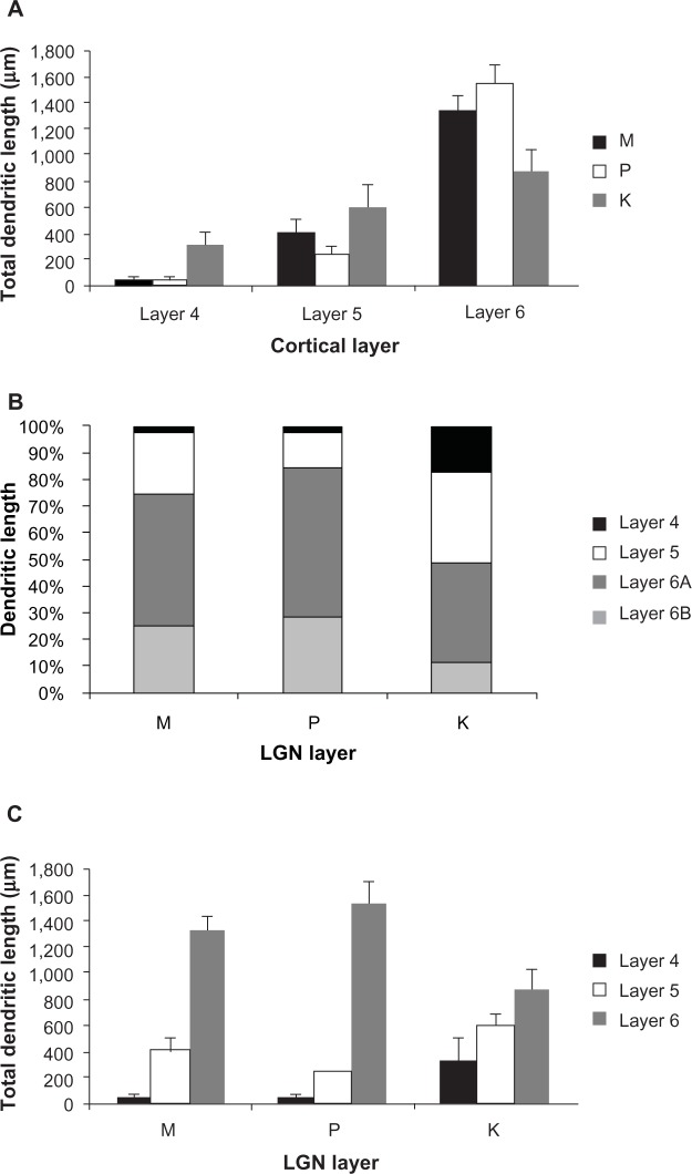
In primates, feedforward visual pathways from retina to lateral geniculate nucleus (LGN) are segregated to different layers. These layers also receive strong reciprocal feedback pathways from cortex. The degree to which feedforward streams in primates are segregated from feedback streams remains unclear. Here, we asked whether corticogeniculate cells that innervate the magnocellular (M), parvocellular (P), and koniocellular (K) layers of the LGN in the prosimian primate bush baby () can be distinguished based on either the laminar distribution or morphological characteristics of their axons and synaptic contacts in LGN, or on their cell body position, size, and dendritic distribution in cortex. Corticogeniculate axons and synapses were labeled anterogradely with biotinylated dextran injections in layer 6 of cortex. Corticogeniculate cell bodies were first labeled with fluorescent dextran injections limited to individual M, P, or K LGN layers and then filled with biotinylated Lucifer yellow. Results showed that feedback to the M or P LGN layers arises from cells with dendrites primarily confined to cortical layer 6 and axons restricted to either M or P LGN layers, but not both. Feedback to K LGN layers arises from cells: 1) whose dendrites distribute rather evenly across cortical layers 5 and 6; 2) whose dendrites always extend into layer 4; and 3) whose axons are never confined to K layers but always overlap with either P or M layers. Corticogeniculate axons also showed distributions that were retinotopically precise based on known receptive field sizes of layer 6 cells, and these axons mainly made synapses with glutamatergic projection neurons in the LGN in all layers. Taken together with prior physiological results, we argue that the morphological differences between the three corticogeniculate pathways show that the M and P feedback pathways could rapidly and specifically enhance local LGN activity, while we speculate that the K feedback pathway is organized to temporally synchronize activity between LGN and cortex.
在灵长类动物中,从视网膜到外侧膝状体核(LGN)的前馈视觉通路被分隔到不同的层。这些层还接受来自皮层的强大的相互反馈通路。灵长类动物的前馈信息流与反馈信息流的分离程度尚不清楚。在这里,我们研究了在原猴灵长类动物婴猴中,支配LGN的大细胞(M)、小细胞(P)和konio细胞(K)层的皮质膝状体细胞,是否可以根据其轴突和在LGN中的突触接触的层状分布或形态特征,或者根据其在皮层中的细胞体位置、大小和树突分布来区分。通过在皮层第6层注射生物素化葡聚糖对皮质膝状体轴突和突触进行顺行标记。首先用限于单个M、P或K LGN层的荧光葡聚糖注射标记皮质膝状体细胞体,然后用生物素化路西法黄填充。结果表明,对M或P LGN层的反馈来自树突主要局限于皮层第6层且轴突限于M或P LGN层之一而非两者的细胞。对K LGN层的反馈来自以下细胞:1)其树突在皮层第5层和第6层中分布相当均匀;2)其树突总是延伸到第4层;3)其轴突从不局限于K层,而是总是与P层或M层重叠。基于第6层细胞已知的感受野大小,皮质膝状体轴突还显示出视网膜拓扑精确的分布,并且这些轴突主要在LGN的所有层中与谷氨酸能投射神经元形成突触。结合先前的生理学结果,我们认为三种皮质膝状体通路之间的形态差异表明,M和P反馈通路可以快速且特异性地增强局部LGN活动,而我们推测K反馈通路的组织方式是使LGN和皮层之间的活动在时间上同步。