Department of Ophthalmology, Miguel Servet University Hospital, Zaragoza, Spain.
Miguel Servet Ophthalmology Research Group (GIMSO), Aragon Health Research Institute (IIS Aragon), University of Zaragoza, Spain.
Invest Ophthalmol Vis Sci. 2021 Oct 4;62(13):9. doi: 10.1167/iovs.62.13.9.
To evaluate differences by sex in the neuroretina of rats with chronic glaucoma over 24 weeks of follow-up, and to assess by sex the influence on neurodegeneration of different methods of inducing ocular hypertension.
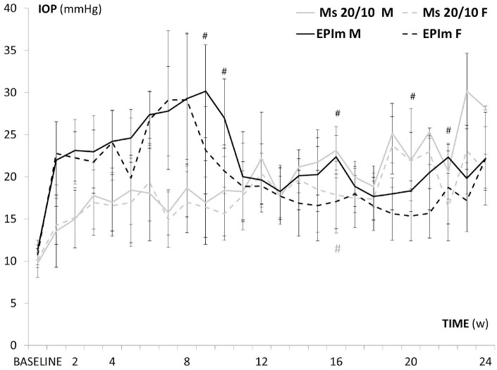
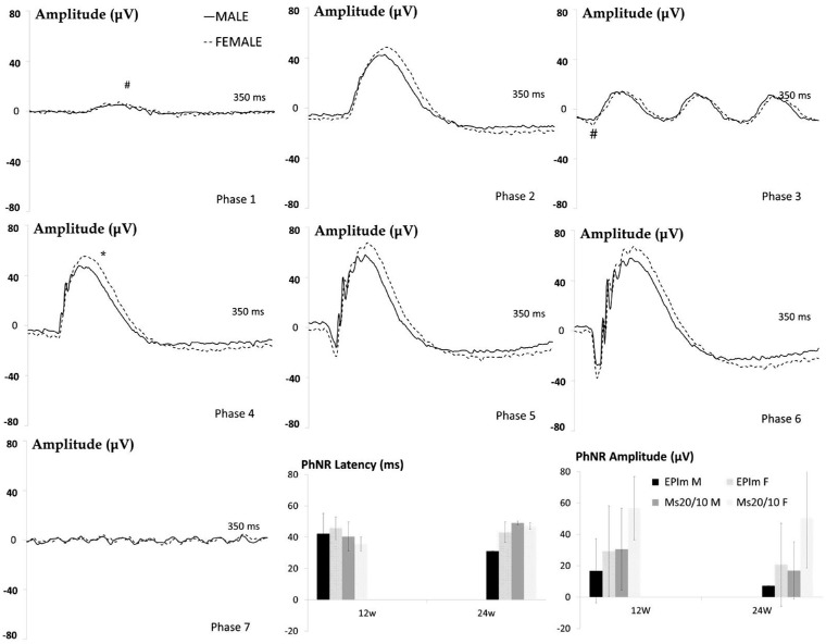
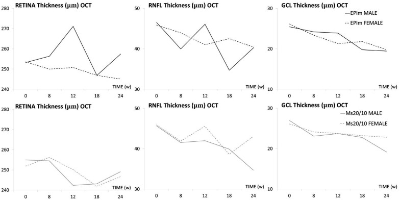
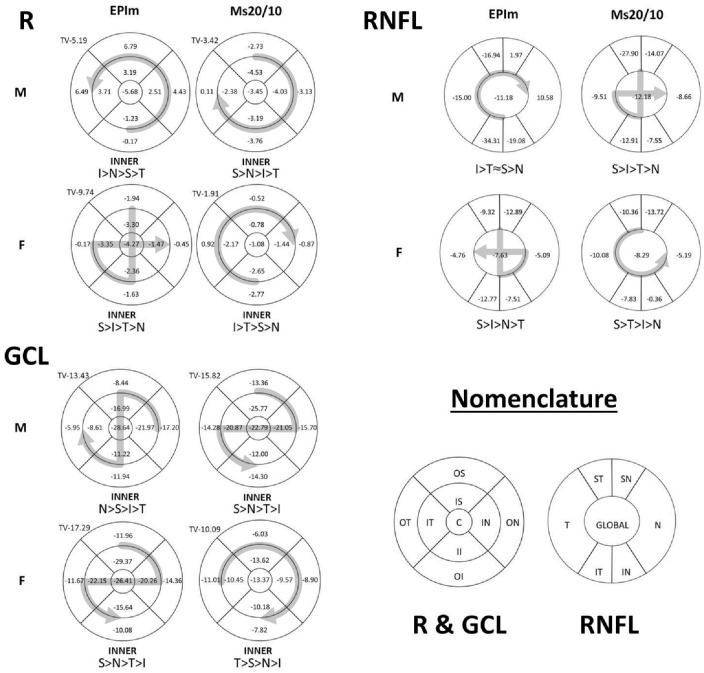
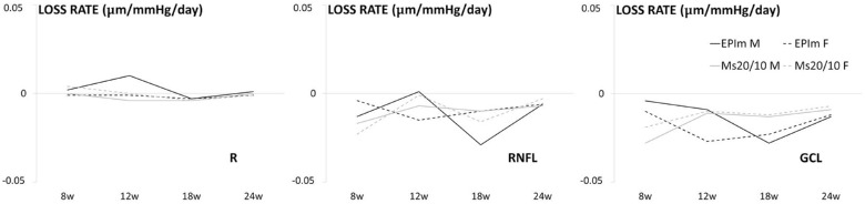
Forty-six Long-Evans rats-18 males and 28 females-with induced chronic glaucoma were analyzed. Glaucoma was achieved via 2 models: repeatedly sclerosing the episcleral veins (9 male/14 female) or by injecting poly(lactic-co-glycolic acid) microspheres measuring 20 to 10 µm (Ms20/10) into the anterior chamber (9 male/14 female). The IOP was measured weekly by tonometer; neuroretinal function was recorded by dark/light-adapted electroretinography at baseline and weeks 12 and 24; and structure was analyzed by optical coherence tomography using the retina posterior pole, retinal nerve fiber layer and ganglion cell layer protocols at baseline and weeks 8, 12, 18, and 24.
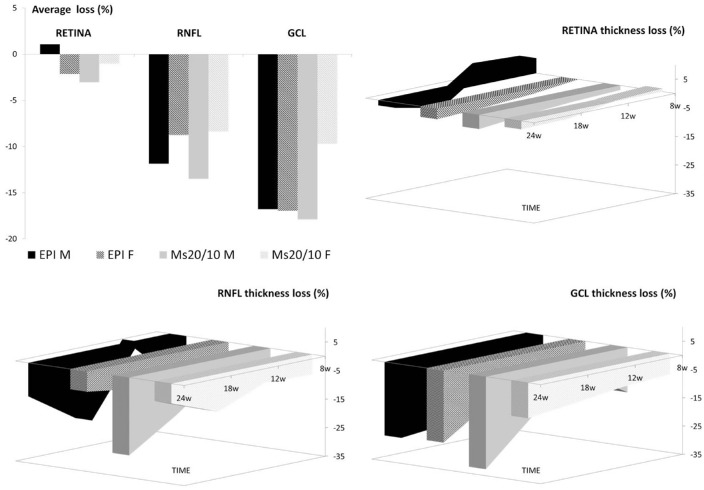
Males showed statistically significant (P < 0.05) higher IOP in both chronic glaucoma models, and greater differences were found in the episcleral model at earlier stages. Males with episclerally induced glaucoma showed a statistically higher increase in retinal thickness in optical coherence tomography recordings than females and also when comparing Ms20/10 at 12 weeks. Males showed a higher percentage of retinal nerve fiber layer thickness loss in both models. Ganglion cell layer thickness loss was only detected in the Ms20/10 model. Males exhibited worse dark/light-adapted functionality in chronic glaucoma models, which worsened in the episcleral sclerosis model at 12 weeks, than females.
Female rats with chronic glaucoma experienced lower IOP and structural loss and better neuroretinal functionality than males. Sex and the ocular hypertension-inducing method influenced neuroretinal degeneration.
评估在 24 周的随访中慢性青光眼大鼠神经视网膜的性别差异,并评估不同诱导眼内高压方法对神经退行性变的影响。
分析了 46 只长耳大鼠(18 只雄性和 28 只雌性)的慢性青光眼。通过两种模型诱导青光眼:反复硬化巩膜静脉(9 只雄性/14 只雌性)或在前房内注射 20 至 10µm 的聚(乳酸-共-乙醇酸)微球(Ms20/10)(9 只雄性/14 只雌性)。每周通过眼压计测量 IOP;在基线和 12 周和 24 周时,通过暗/亮适应视网膜电图记录神经视网膜功能;在基线和第 8、12、18 和 24 周时,通过光学相干断层扫描使用视网膜后极、视网膜神经纤维层和节细胞层方案分析结构。
两种慢性青光眼模型中,雄性的 IOP 均显著升高(P < 0.05),在前节巩膜模型中早期差异更大。前节巩膜诱导青光眼的雄性在光学相干断层扫描记录中的视网膜厚度增加比雌性更高,与 Ms20/10 在 12 周时相比也更高。两种模型中,雄性的视网膜神经纤维层厚度损失百分比均较高。仅在 Ms20/10 模型中检测到节细胞层厚度损失。在慢性青光眼模型中,雄性的暗/亮适应功能比雌性更差,在前节巩膜硬化模型中 12 周时恶化。
慢性青光眼的雌性大鼠经历的眼压和结构损失较低,神经视网膜功能更好。性别和诱导眼内高压的方法影响神经视网膜变性。