Julius Wolff Institute, Berlin Institute of Health at Charité - Universitätsmedizin Berlin, 13353, Berlin, Germany.
BIH-Center for Regenerative Therapies, Berlin Institute of Health at Charité - Universitätsmedizin Berlin, 13353, Berlin, Germany.
BMC Musculoskelet Disord. 2021 Oct 14;22(1):877. doi: 10.1186/s12891-021-04752-1.
The role of the subacromial bursa in the development or healing of shoulder pathologies is unclear. Due to this limited knowledge, we aimed to understand specific reactions of the subacromial bursa according to rotator cuff (RC) pathologies compared to non-tendon defects of the shoulder. We hypothesized that the tissue composition and inflammatory status of the bursa are likely to vary between shoulder pathologies depending on the presence and the extent of RC lesion.
Bursa samples from patients with either 1) shoulder instability with intact RC (healthy bursa, control), 2) osteochondral pathology with intact RC, 3) partial supraspinatus (SSP) tendon tear, or 4) full-thickness SSP tear were investigated histologically and on gene expression level.
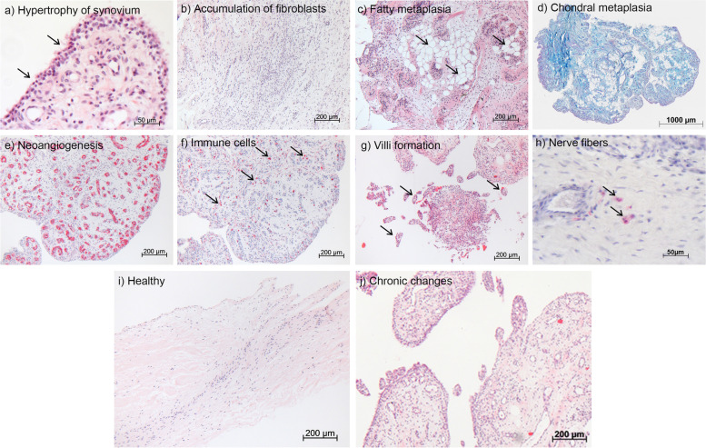
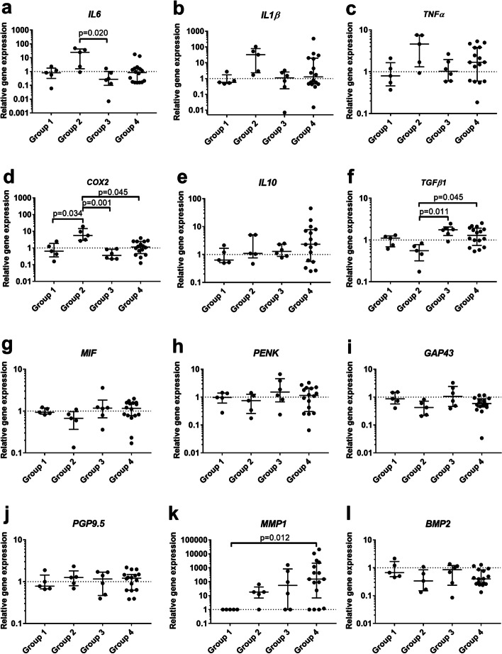
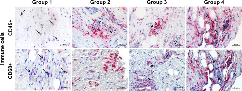
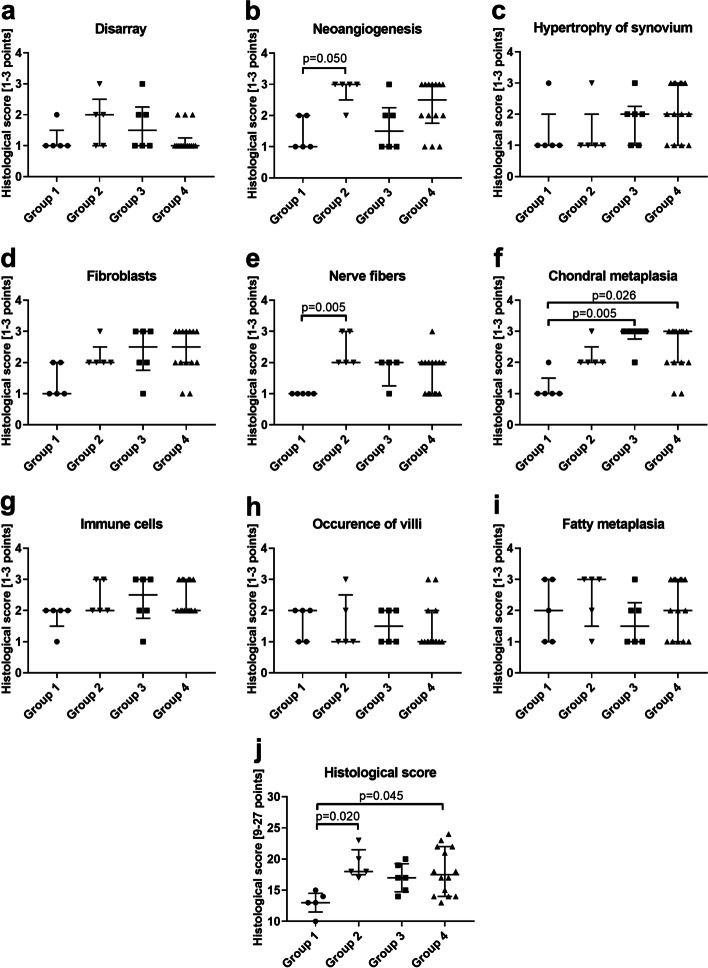
Bursae from SSP tears differed from non-tendon pathologies by exhibiting increased chondral metaplasia and TGFβ1 expression. MMP1 was not expressed in healthy bursa controls, but strongly increased with full-thickness SSP tears. Additionally, the expression of the inflammatory mediators IL1β, IL6, and COX2 increased with the extent of SSP tear as shown by correlation analysis. In contrast, increased angiogenesis and nerve fibers as well as significantly upregulated IL6 and COX2 expression were features of bursae from patients with osteochondral pathology. Using immunohistochemistry, CD45+ leukocytes were observed in all examined groups, which were identified in particular as CD68+ monocytes/macrophages.
In summary, besides the strong increase in MMP1 expression with SSP tear, molecular changes were minor between the investigated groups. However, expression of pro-inflammatory cytokines correlated with the severity of the SSP tear. Most pronounced tissue alterations occurred for the osteochondral pathology and full-thickness SSP tear group, which demonstrates that the bursal reaction is not exclusively dependent on the occurrence of an SSP tear rather than longstanding degenerative changes. The present bursa characterization contributes to the understanding of specific tissue alterations related to RC tears or non-tendon shoulder pathologies. This pilot study provides the basis for future studies elucidating the role of the subacromial bursa in the development or healing of shoulder pathologies.
肩峰下囊在肩部病变的发展或愈合中的作用尚不清楚。由于这方面知识有限,我们旨在了解肩袖(RC)病变与非肌腱肩损伤相比,肩峰下囊的特定反应。我们假设,根据 RC 损伤的存在和程度,囊的组织成分和炎症状态可能会因肩部病变而异。
研究了来自以下患者的囊样本:1)肩不稳伴 RC 完整(健康囊,对照组),2)骨软骨病变伴 RC 完整,3)部分冈上肌腱撕裂,或 4)全层冈上肌腱撕裂。对其进行组织学和基因表达水平分析。
与非肌腱病变相比,冈上肌腱撕裂的囊表现出增加的软骨化生和 TGFβ1 表达。MMP1 在健康囊对照中不表达,但在全层冈上肌腱撕裂中强烈增加。此外,通过相关性分析,随着冈上肌腱撕裂程度的增加,炎症介质 IL1β、IL6 和 COX2 的表达也增加。相比之下,骨软骨病变患者的囊中观察到增加的血管生成和神经纤维以及显著上调的 IL6 和 COX2 表达。使用免疫组织化学,在所有检查的组中均观察到 CD45+白细胞,特别是 CD68+单核细胞/巨噬细胞。
总之,除了冈上肌腱撕裂时 MMP1 表达的强烈增加外,在研究组之间,分子变化较小。然而,促炎细胞因子的表达与冈上肌腱撕裂的严重程度相关。最明显的组织改变发生在骨软骨病变和全层冈上肌腱撕裂组,这表明囊的反应不仅取决于冈上肌腱撕裂的发生,还取决于长期的退行性变化。本研究为理解与 RC 撕裂或非肌腱肩部病变相关的特定组织改变提供了基础。这项初步研究为阐明肩峰下囊在肩部病变发展或愈合中的作用提供了依据。