Radiology and Medical Imaging Department, College of Applied Medical Sciences, Prince Sattam Bin Abdulaziz University, PO Box 422, Al-Kharj, 11942, Saudi Arabia.
Medical Imaging Department, Prince Mohammed Bin Abdulaziz Hospital, Riyadh, Saudi Arabia.
Sci Rep. 2021 Oct 14;11(1):20476. doi: 10.1038/s41598-021-00064-5.
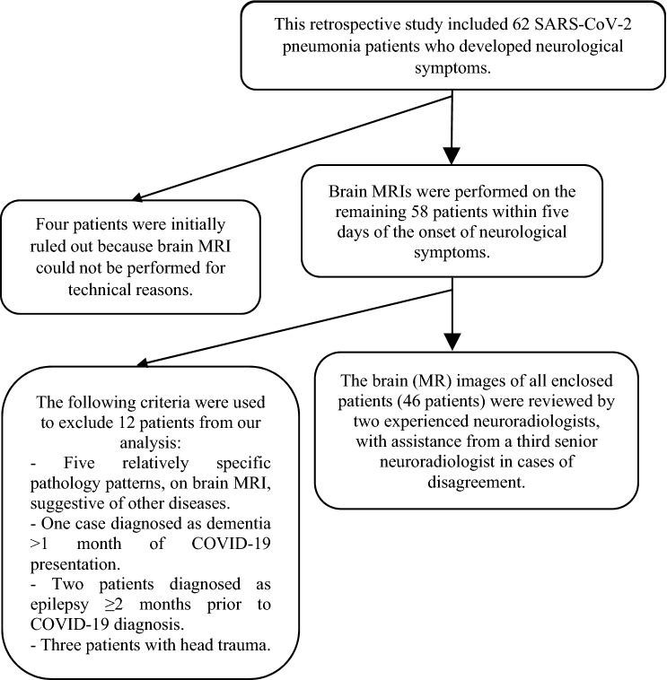
The increased frequency of neurological manifestations, including central nervous system (CNS) manifestations, in patients with coronavirus disease 2019 (COVID-19) pandemic is consistent with the virus's neurotropic nature. In most patients, brain magnetic resonance imaging (MRI) is a sensitive imaging modality in the diagnosis of viral encephalitides in the brain. The purpose of this study was to determine the frequency of brain lesion patterns on brain MRI in severe acute respiratory syndrome coronavirus 2 (SARS-CoV-2) pneumonia patients who developed focal and non-focal neurological manifestations. In addition, it will compare the impact of the Glasgow Coma Scale (GCS) as an index of deteriorating cerebral function on positive brain MRIs in both neurological manifestations. This retrospective study included an examination of SARS-CoV-2 pneumonia patients with real-time reverse transcription polymerase chain reaction (RT-PCR) confirmation, admitted with clinicoradiologic evidence of COVID-19 pneumonia, and who were candidates for brain MRI due to neurological manifestations suggesting brain involvement. Brain imaging acquired on a 3.0 T MRI system (Skyra; Siemens, Erlangen, Germany) with a 20-channel receive head coil. Brain MRI revealed lesions in 38 (82.6%) of the total 46 patients for analysis and was negative in the remaining eight (17.4%) of all finally enclosed patients with RT-PCR confirmed SARS-CoV-2 pneumonia. Twenty-nine (63%) patients had focal neurological manifestations, while the remaining 17 (37%) patients had non-focal neurological manifestations. The patients had a highly significant difference (p = 0.0006) in GCS, but no significant difference (p = 0.4) in the number of comorbidities they had. Brain MRI is a feasible and important imaging modality in patients with SARS-CoV-2 pneumonia who develop neurological manifestations suggestive of brain involvement, particularly in patients with non-focal manifestations and a decline in GCS.
新型冠状病毒病 2019(COVID-19)大流行中,神经系统表现(包括中枢神经系统表现)的频率增加与病毒的嗜神经性一致。在大多数患者中,脑磁共振成像(MRI)是诊断脑部病毒性脑炎的一种敏感影像学方法。本研究的目的是确定发生局灶性和非局灶性神经表现的严重急性呼吸综合征冠状病毒 2(SARS-CoV-2)肺炎患者脑 MRI 上脑部病变模式的频率。此外,它将比较格拉斯哥昏迷量表(GCS)作为脑功能恶化指标对两种神经表现中阳性脑 MRI 的影响。这项回顾性研究包括对实时逆转录聚合酶链反应(RT-PCR)确认的 SARS-CoV-2 肺炎患者进行检查,这些患者具有临床放射学证据表明患有 COVID-19 肺炎,并且由于提示脑部受累的神经表现而适合进行脑部 MRI。脑成像在 3.0 T MRI 系统(Skyra;西门子,德国埃朗根)上进行,使用 20 通道接收头部线圈。脑 MRI 显示 46 名患者中有 38 名(82.6%)的总患者有病变,而在所有最终包含 RT-PCR 确认 SARS-CoV-2 肺炎的患者中,其余 8 名(17.4%)患者的脑 MRI 为阴性。29 名(63%)患者有局灶性神经表现,而其余 17 名(37%)患者有非局灶性神经表现。患者的 GCS 差异有统计学意义(p=0.0006),但并发症数量无统计学差异(p=0.4)。脑 MRI 是一种可行且重要的影像学方法,适用于发生提示脑部受累的神经表现的 SARS-CoV-2 肺炎患者,特别是在 GCS 下降的非局灶性表现患者中。