Zare Dehnavi Ali, Salehi Mohammadreza, Arab Ahmadi Mehran, Asgardoon Mohammad Hossein, Ashrafi Farzad, Ahmadinejad Nasrin, Behkar Atefeh, Hamidi Farahani Ramin, Hashemi Hassan, Tafakhori Abbas, Shahali Hamze, Rahmani Mohammad, Ranjbar Naeini Alireza
Department of Neurology, School of Medicine, AJA University of Medical Sciences, Tehran, Iran.
Infectious Diseases and Tropical Medicines Department, Tehran University of Medical Sciences, Tehran, Iran.
Arch Acad Emerg Med. 2022 Jan 30;10(1):e10. doi: 10.22037/aaem.v10i1.1507. eCollection 2022.
Although neurologic involvement and neuroimaging abnormalities have been frequently identified in COVID-19 patients, the underlying factors remain unclear. In this study, we assessed the association of the neurological manifestations and neuroimaging features of hospitalized COVID-19 patients with their clinical, laboratory, and imaging characteristics.
This multicenter cross-sectional study was conducted between September 2020 and March 2021 at two large academic hospitals in Tehran, Iran. We used census sampling from medical records to enroll hospitalized patients with a positive COVID-19 Polymerase chain reaction (PCR) test who underwent brain imaging due to presenting any acute neurologic symptom during hospital stay.
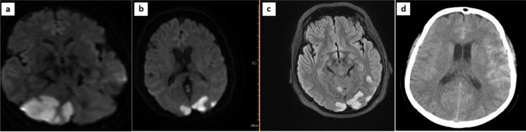
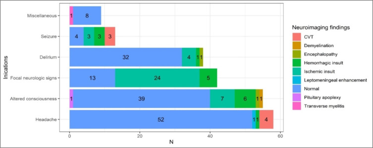
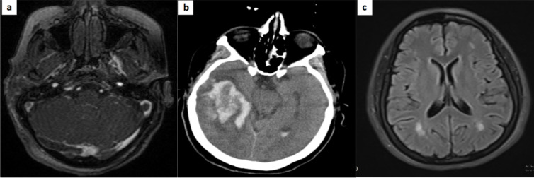
Of the 4372 hospitalized patients with COVID-19, only 211 met the inclusion criteria (35.5% with severe infection). Central nervous system and psychiatric manifestations were significantly more common in severe cases (p ≤ 0.044). Approximately, 30% had a new abnormality on their neuroimaging, with ischemic (38/63) and hemorrhagic (16/63) insults being the most common. The most frequent reasons that provoked cranial imaging were headache (27%), altered consciousness (25.6%), focal neurologic signs (19.9%), and delirium (18%). Analysis revealed a positive correlation for age, neutrophilia, lymphopenia, erythrocyte sedimentation rate (ESR), and C-reactive protein (CRP) with the emergence of neuroimaging abnormalities (p ≤ 0.018). In addition, patients with new neuroimaging abnormalities had a significantly higher lung CT score than those without any pathologic findings (11.1 ± 4.8 vs. 5.9 ± 4.8, p < 0.001).
Approximately 30% of the study population had various acute neuroimaging findings. The lung CT score, neutrophil count, and age were strong predictors of acute neuroimaging abnormalities in hospitalized COVID-19 patients.
尽管在新型冠状病毒肺炎(COVID-19)患者中经常发现神经系统受累及神经影像学异常,但其潜在因素仍不清楚。在本研究中,我们评估了住院COVID-19患者的神经学表现和神经影像学特征与其临床、实验室及影像学特征之间的关联。
本多中心横断面研究于2020年9月至2021年3月在伊朗德黑兰的两家大型学术医院进行。我们从病历中采用普查抽样的方法,纳入因住院期间出现任何急性神经症状而接受脑部成像检查且COVID-19聚合酶链反应(PCR)检测呈阳性的住院患者。
在4372例住院COVID-19患者中,只有211例符合纳入标准(35.5%为重症感染)。中枢神经系统和精神症状在重症病例中明显更为常见(p≤0.044)。约30%的患者神经影像学出现新的异常,其中缺血性(38/63)和出血性(16/63)损伤最为常见。引发头颅成像检查的最常见原因是头痛(27%)、意识改变(25.6%)、局灶性神经体征(19.9%)和谵妄(18%)。分析显示年龄、中性粒细胞增多、淋巴细胞减少、红细胞沉降率(ESR)和C反应蛋白(CRP)与神经影像学异常的出现呈正相关(p≤0.018)。此外,神经影像学有新异常的患者肺部CT评分显著高于无任何病理发现的患者(11.1±4.8 vs. 5.9±4.8,p<0.001)。
约30%的研究人群有各种急性神经影像学表现。肺部CT评分、中性粒细胞计数和年龄是住院COVID-19患者急性神经影像学异常的有力预测指标。