• 文献检索
  • 文档翻译
  • 深度研究
  • 学术资讯
  • Suppr Zotero 插件Zotero 插件
  • 邀请有礼
  • 套餐&价格
  • 历史记录
应用&插件
Suppr Zotero 插件Zotero 插件浏览器插件Mac 客户端Windows 客户端微信小程序
定价
高级版会员购买积分包购买API积分包
服务
文献检索文档翻译深度研究API 文档MCP 服务
关于我们
关于 Suppr公司介绍联系我们用户协议隐私条款
关注我们

Suppr 超能文献

核心技术专利:CN118964589B侵权必究
粤ICP备2023148730 号-1Suppr @ 2026

文献检索

告别复杂PubMed语法,用中文像聊天一样搜索,搜遍4000万医学文献。AI智能推荐,让科研检索更轻松。

立即免费搜索

文件翻译

保留排版,准确专业,支持PDF/Word/PPT等文件格式,支持 12+语言互译。

免费翻译文档

深度研究

AI帮你快速写综述,25分钟生成高质量综述,智能提取关键信息,辅助科研写作。

立即免费体验

乳腺癌钙化:一种新分割方法的识别。

Breast Cancer Calcifications: Identification Using a Novel Segmentation Approach.

机构信息

University of Engineering and Management, Kolkata, India.

School of Computer Science and Engineering, Lovely Professional University, Phagwara, Punjab, India.

出版信息

Comput Math Methods Med. 2021 Oct 6;2021:9905808. doi: 10.1155/2021/9905808. eCollection 2021.

DOI:10.1155/2021/9905808
PMID:34659451
原文链接:https://pmc.ncbi.nlm.nih.gov/articles/PMC8514925/
Abstract

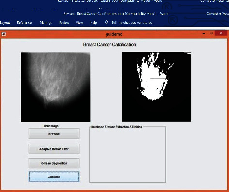
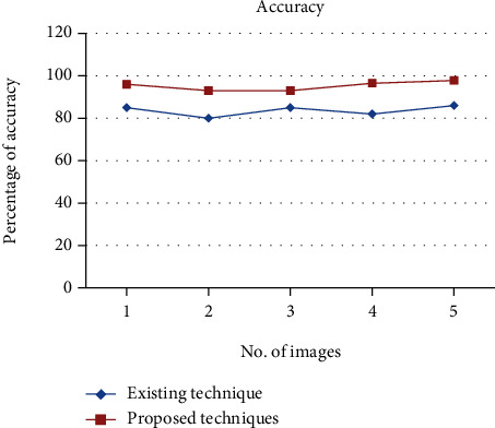
Breast cancer is a strong risk factor of cancer amongst women. One in eight women suffers from breast cancer. It is a life-threatening illness and is utterly dreadful. The root cause which is the breast cancer agent is still under research. There are, however, certain potentially dangerous factors like age, genetics, obesity, birth control, cigarettes, and tablets. Breast cancer is often a malignant tumor that begins in the breast cells and eventually spreads to the surrounding tissue. If detected early, the illness may be reversible. The probability of preservation diminishes as the number of measurements increases. Numerous imaging techniques are used to identify breast cancer. This research examines different breast cancer detection strategies via the use of imaging techniques, data mining techniques, and various characteristics, as well as a brief comparative analysis of the existing breast cancer detection system. Breast cancer mortality will be significantly reduced if it is identified and treated early. There are technological difficulties linked to scans and people's inconsistency with breast cancer. In this study, we introduced a form of breast cancer diagnosis. There are different methods involved to collect and analyze details. In the preprocessing stage, the input data picture is filtered by using a window or by cropping. Segmentation can be performed using -means algorithm. This study is aimed at identifying the calcifications found in bosom cancer in the last phase. The suggested approach is already implemented in MATLAB, and it produces reliable performance.

摘要

乳腺癌是女性癌症的一个强烈危险因素。每 8 个女性中就有 1 个患有乳腺癌。这是一种危及生命的疾病,非常可怕。导致乳腺癌的根本原因仍在研究中。然而,有一些潜在的危险因素,如年龄、遗传、肥胖、避孕药、香烟和药片。乳腺癌通常是一种恶性肿瘤,始于乳腺细胞,最终扩散到周围组织。如果早期发现,疾病可能是可逆的。随着测量次数的增加,保存的可能性会降低。有许多成像技术可用于识别乳腺癌。这项研究通过使用成像技术、数据挖掘技术和各种特征,以及对现有的乳腺癌检测系统进行简要的比较分析,探讨了不同的乳腺癌检测策略。如果早期发现并治疗,乳腺癌的死亡率将显著降低。扫描技术存在困难,而且人们对乳腺癌的认识不一致。在这项研究中,我们引入了一种乳腺癌诊断方法。有不同的方法涉及到收集和分析细节。在预处理阶段,使用窗口或裁剪对输入数据图像进行滤波。可以使用 -means 算法进行分割。本研究旨在识别乳腺癌症后期发现的钙化。该方法已经在 MATLAB 中实现,并且产生了可靠的性能。

https://cdn.ncbi.nlm.nih.gov/pmc/blobs/a9cc/8514925/ad89269c9736/CMMM2021-9905808.008.jpg
https://cdn.ncbi.nlm.nih.gov/pmc/blobs/a9cc/8514925/c4dc40281747/CMMM2021-9905808.001.jpg
https://cdn.ncbi.nlm.nih.gov/pmc/blobs/a9cc/8514925/f2087da1a026/CMMM2021-9905808.002.jpg
https://cdn.ncbi.nlm.nih.gov/pmc/blobs/a9cc/8514925/dbe6f279a867/CMMM2021-9905808.003.jpg
https://cdn.ncbi.nlm.nih.gov/pmc/blobs/a9cc/8514925/f1fb3cae8c7f/CMMM2021-9905808.004.jpg
https://cdn.ncbi.nlm.nih.gov/pmc/blobs/a9cc/8514925/c60629e0b6b6/CMMM2021-9905808.005.jpg
https://cdn.ncbi.nlm.nih.gov/pmc/blobs/a9cc/8514925/0917f70dff37/CMMM2021-9905808.006.jpg
https://cdn.ncbi.nlm.nih.gov/pmc/blobs/a9cc/8514925/38b385c63c49/CMMM2021-9905808.007.jpg
https://cdn.ncbi.nlm.nih.gov/pmc/blobs/a9cc/8514925/ad89269c9736/CMMM2021-9905808.008.jpg

相似文献

1
Breast Cancer Calcifications: Identification Using a Novel Segmentation Approach.乳腺癌钙化:一种新分割方法的识别。
Comput Math Methods Med. 2021 Oct 6;2021:9905808. doi: 10.1155/2021/9905808. eCollection 2021.
2
Differentiating Grade in Breast Invasive Ductal Carcinoma Using Texture Analysis of MRI.基于 MRI 纹理分析鉴别乳腺浸润性导管癌分级。
Comput Math Methods Med. 2020 Apr 7;2020:6913418. doi: 10.1155/2020/6913418. eCollection 2020.
3
A Novel Internet of Things Framework Integrated with Real Time Monitoring for Intelligent Healthcare Environment.一种新型物联网框架,集成实时监测,用于智能医疗保健环境。
J Med Syst. 2019 May 3;43(6):165. doi: 10.1007/s10916-019-1302-9.
4
[Computer aided diagnosis of calcifications in mammograms].[乳腺X线摄影中钙化的计算机辅助诊断]
Sheng Wu Yi Xue Gong Cheng Xue Za Zhi. 2011 Feb;28(1):170-4.
5
Deep feature-based automatic classification of mammograms.基于深度特征的乳腺X线照片自动分类
Med Biol Eng Comput. 2020 Jun;58(6):1199-1211. doi: 10.1007/s11517-020-02150-8. Epub 2020 Mar 21.
6
Improving the Mann-Whitney statistical test for feature selection: an approach in breast cancer diagnosis on mammography.改进用于特征选择的曼-惠特尼统计检验:一种乳腺钼靶摄影乳腺癌诊断方法
Artif Intell Med. 2015 Jan;63(1):19-31. doi: 10.1016/j.artmed.2014.12.004. Epub 2014 Dec 12.
7
Computer aided detection of microcalcifications in digital mammograms.数字乳腺钼靶片中微钙化的计算机辅助检测
Comput Biol Med. 2000 Sep;30(5):267-86. doi: 10.1016/s0010-4825(00)00014-7.
8
Computer aided detection system for micro calcifications in digital mammograms.数字乳腺钼靶片中微钙化的计算机辅助检测系统
Comput Methods Programs Biomed. 2014 Oct;116(3):226-35. doi: 10.1016/j.cmpb.2014.04.010. Epub 2014 Apr 30.
9
Computerized segmentation method for individual calcifications within clustered microcalcifications while maintaining their shapes on magnification mammograms.基于放大数字化乳腺图像的簇状微钙化中个体钙化的计算机分割方法,同时保持其形态。
J Digit Imaging. 2012 Jun;25(3):377-86. doi: 10.1007/s10278-011-9420-z.
10
An Improved Brain MRI Classification Methodology Based on Statistical Features and Machine Learning Algorithms.基于统计特征和机器学习算法的改进型脑 MRI 分类方法。
Comput Math Methods Med. 2021 Dec 7;2021:8608305. doi: 10.1155/2021/8608305. eCollection 2021.

引用本文的文献

1
An enhanced denoising system for mammogram images using deep transformer model with fusion of local and global features.一种使用具有局部和全局特征融合的深度变压器模型的乳腺X光图像增强去噪系统。
Sci Rep. 2025 Feb 24;15(1):6562. doi: 10.1038/s41598-025-89451-w.
2
A Lightweight Hybrid Dilated Ghost Model-Based Approach for the Prognosis of Breast Cancer.一种基于轻量级混合扩张式幽灵模型的乳腺癌预后方法
Comput Intell Neurosci. 2022 Aug 25;2022:9325452. doi: 10.1155/2022/9325452. eCollection 2022.
3
Retracted: Breast Cancer Calcifications: Identification Using a Novel Segmentation Approach.

本文引用的文献

1
Prediction of Heart Disease Using a Combination of Machine Learning and Deep Learning.基于机器学习和深度学习的心脏病预测。
Comput Intell Neurosci. 2021 Jul 1;2021:8387680. doi: 10.1155/2021/8387680. eCollection 2021.
2
Chest CT Findings and Differential Diagnosis of Pneumonia and Combined with Streptococcal Pneumonia in Children.儿童肺炎与链球菌肺炎的胸部 CT 表现及鉴别诊断。
J Healthc Eng. 2021 Jun 14;2021:8085530. doi: 10.1155/2021/8085530. eCollection 2021.
3
DU-Net: Convolutional Network for the Detection of Arterial Calcifications in Mammograms.
撤回:乳腺癌钙化:使用一种新型分割方法进行识别。
Comput Math Methods Med. 2023 Jul 19;2023:9891703. doi: 10.1155/2023/9891703. eCollection 2023.
4
Face mask recognition system using CNN model.使用卷积神经网络(CNN)模型的口罩识别系统。
Neurosci Inform. 2022 Sep;2(3):100035. doi: 10.1016/j.neuri.2021.100035. Epub 2021 Dec 9.
5
Monitoring Cardiovascular Problems in Heart Patients Using Machine Learning.使用机器学习监测心脏病患者的心血管问题。
J Healthc Eng. 2023 Feb 8;2023:9738123. doi: 10.1155/2023/9738123. eCollection 2023.
6
Visual bias could impede diagnostic accuracy of breast cancer calcifications.视觉偏差可能会妨碍乳腺癌钙化的诊断准确性。
J Med Imaging (Bellingham). 2022 May;9(3):035503. doi: 10.1117/1.JMI.9.3.035503. Epub 2022 Jun 9.
7
Effective Image Processing and Segmentation-Based Machine Learning Techniques for Diagnosis of Breast Cancer.基于有效图像处理和分割的机器学习技术在乳腺癌诊断中的应用。
Comput Math Methods Med. 2022 Apr 8;2022:6841334. doi: 10.1155/2022/6841334. eCollection 2022.
DU-Net:用于乳腺钼靶片中动脉钙化检测的卷积网络。
IEEE Trans Med Imaging. 2020 Oct;39(10):3240-3249. doi: 10.1109/TMI.2020.2989737. Epub 2020 Apr 23.
4
Breast Calcifications Detection Based on Radiofrequency Signals by Quantitative Ultrasound Multi-parameter Fusion.基于定量超声多参数融合的射频信号乳腺钙化检测
Annu Int Conf IEEE Eng Med Biol Soc. 2019 Jul;2019:6196-6199. doi: 10.1109/EMBC.2019.8857908.
5
Breast Microcalcification Diagnosis Using Deep Convolutional Neural Network from Digital Mammograms.基于数字乳腺 X 线摄影的深度学习卷积神经网络在乳腺微钙化中的诊断应用
Comput Math Methods Med. 2019 Mar 3;2019:2717454. doi: 10.1155/2019/2717454. eCollection 2019.
6
Analysis of the Cluster Prominence Feature for Detecting Calcifications in Mammograms.乳腺 X 线摄影中簇状突出特征分析在检测钙化中的应用
J Healthc Eng. 2018 Dec 30;2018:2849567. doi: 10.1155/2018/2849567. eCollection 2018.
7
Improving the Automated Detection of Calcifications Using Adaptive Variance Stabilization.利用自适应方差稳定化提高钙化点的自动检测效果。
IEEE Trans Med Imaging. 2018 Aug;37(8):1857-1864. doi: 10.1109/TMI.2018.2814058. Epub 2018 Mar 9.
8
A hierarchical pipeline for breast boundary segmentation and calcification detection in mammograms.一种用于乳腺边界分割和乳腺 X 线片中钙化检测的分层流水线。
Comput Biol Med. 2018 May 1;96:178-188. doi: 10.1016/j.compbiomed.2018.03.011. Epub 2018 Mar 16.
9
Ultrasound Imaging Technologies for Breast Cancer Detection and Management: A Review.用于乳腺癌检测与管理的超声成像技术:综述
Ultrasound Med Biol. 2018 Jan;44(1):37-70. doi: 10.1016/j.ultrasmedbio.2017.09.012. Epub 2017 Oct 26.
10
Automated Analysis of Unregistered Multi-View Mammograms With Deep Learning.基于深度学习的未注册多视图乳腺 X 光片的自动分析。
IEEE Trans Med Imaging. 2017 Nov;36(11):2355-2365. doi: 10.1109/TMI.2017.2751523. Epub 2017 Sep 12.