Research and Development Department, MED-EL Medical Electronics, Fürstenweg77a, 6020, Innsbruck, Austria.
Department of Translational Neurosciences, Faculty of Medicine and Health Sciences, University of Antwerp, Antwerp, Belgium.
Sci Rep. 2021 Oct 21;11(1):20868. doi: 10.1038/s41598-021-00330-6.
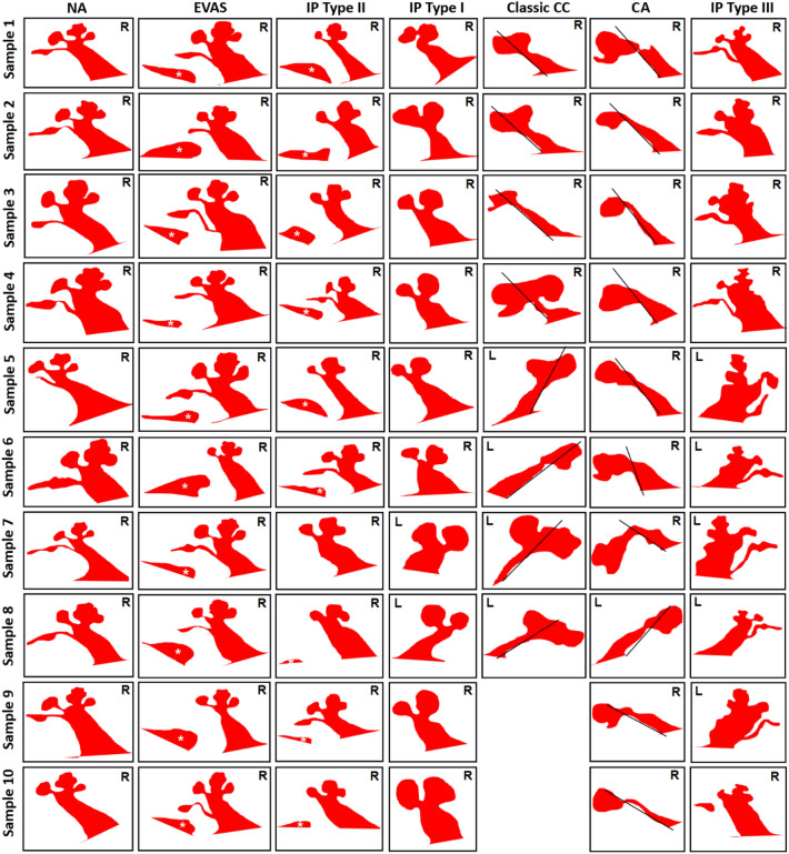
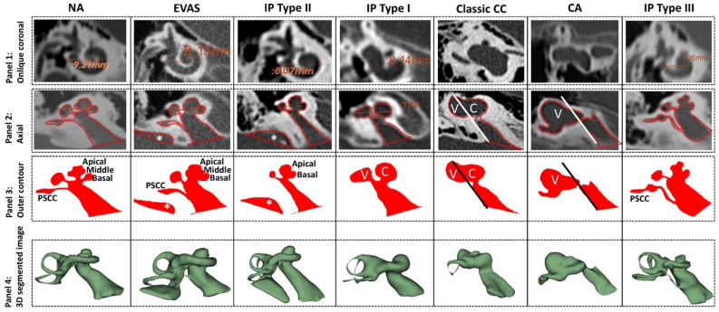
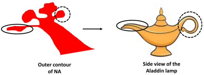
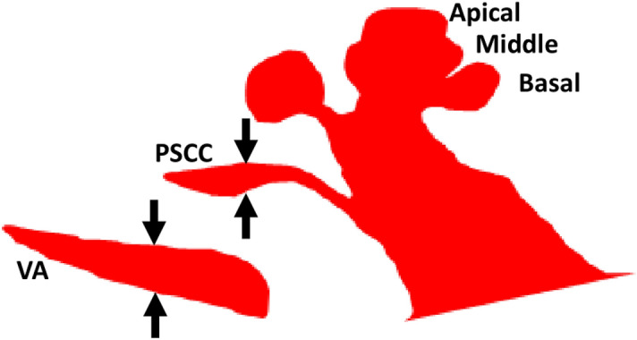
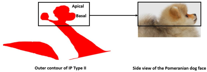
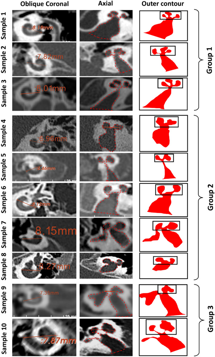
Identification of the inner ear malformation types from radiographs is a complex process. We hypothesize that each inner ear anatomical type has a uniqueness in its appearance in radiographs. The outer contour of the inner ear was captured from the mid-modiolar section, perpendicular to the oblique-coronal plane, from which the A-value was determined from CT scans with different inner ear anatomical types. The mean A-value of normal anatomy (NA) and enlarged vestibular aqueduct syndrome (EVAS) anatomical types was greater than for Incomplete Partition (IP) type I, II, III and cochlear hypoplasia. The outer contour of the cochlear portion within the mid-modiolar section of NA and EVAS resembles the side view of Aladdin's lamp; IP type I resembles the side-view of the Sphinx pyramid and type II a Pomeranian dog's face. The steep spiraling cochlear turns of IP type III resemble an Auger screw tip. Drawing a line parallel to the posterior margin of internal auditory canal (IAC) in axial-view, bisecting the cavity into cochlear and vestibular portions, identifies common-cavity; whereas a cavity that falls under the straight-line leaving no cochlear portion identifies cochlear aplasia. An atlas of the outer contour of seventy-eight inner ears was created for the identification of the inner malformation types precisely.
从影像学上识别内耳畸形类型是一个复杂的过程。我们假设每个内耳解剖类型在影像学上都有其独特的表现。从垂直于斜冠状面的中耳蜗轴位片上获取内耳的外轮廓,并从 CT 扫描中确定 A 值,这些 CT 扫描具有不同的内耳解剖类型。正常解剖(NA)和扩大前庭水管综合征(EVAS)解剖类型的平均 A 值大于不完全分隔(IP)I 型、II 型、III 型和耳蜗发育不全。NA 和 EVAS 中耳蜗部分的中耳蜗轴位片的外轮廓类似于阿拉丁神灯的侧视图;IP 型 I 类似于狮身人面像的侧视图,II 型类似于博美犬的脸。IP 型 III 的陡峭螺旋耳蜗转弯类似于螺旋钻尖端。在轴位视图中,平行于内听道(IAC)后缘画一条线,将腔分为耳蜗和前庭部分,可识别共同腔;而直线下方没有耳蜗部分的腔则可识别为耳蜗发育不全。为了准确识别内耳畸形类型,我们创建了 78 个内耳外轮廓图谱。