MED-EL GmbH, Innsbruck, Austria.
Anat Rec (Hoboken). 2019 Oct;302(10):1792-1799. doi: 10.1002/ar.24136. Epub 2019 Apr 24.
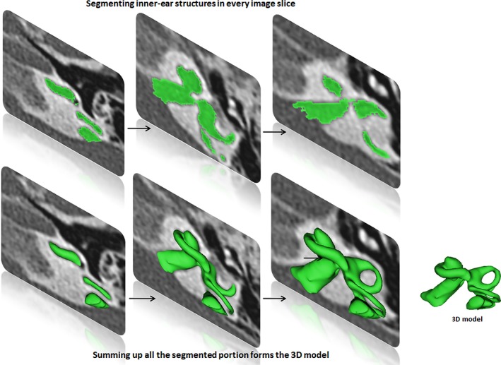
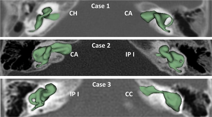
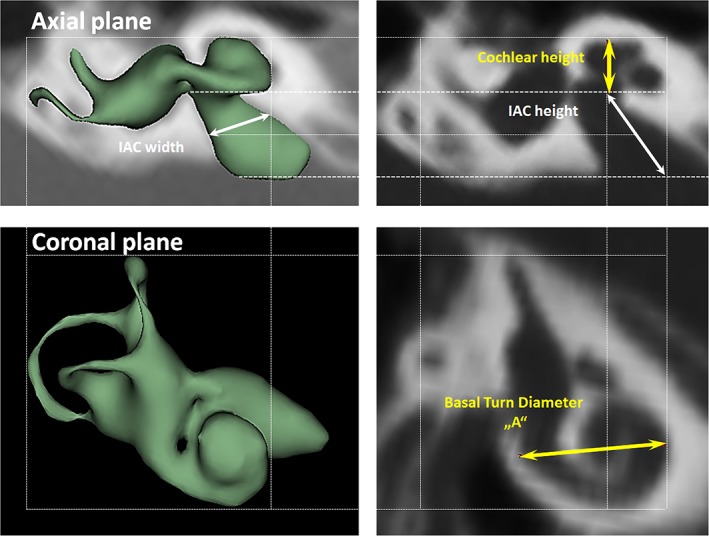
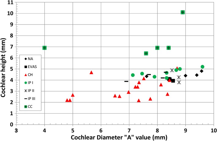
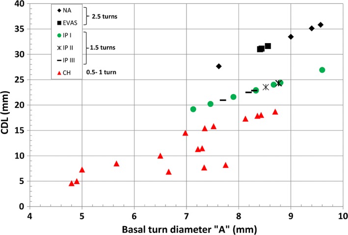
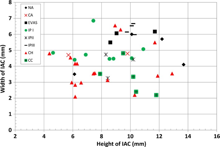
The objective of this study is to determine the variations in size and shape of the most widely recognized cochlear malformation types using three-dimensional (3D) visualization. Using 3D slicer freeware, the complete inner-ear structures were segmented from 46 anonymized high-resolution computed tomography (HRCT) image datasets. Cochlear height, internal auditory canal height, and width were measured from the axial plane. Cochlear basal turn diameter was measured from the oblique coronal plane. Number of cochlear turns was measured from the 3D images and the corresponding cochlear duct length (CDL) was estimated using the CDL equations given in Alexiades et al. [Otol Neurotol 36 (2015) 904-907]. Out of 46 preoperative HRCT image datasets of human temporal bone, cochlear anatomy types including normal anatomy (4), enlarged vestibular aqueduct syndrome (3), cochlear aplasia (2), incomplete partition Types I (8), II (Mondini's deformity) (3), and III (X-linked) (4), cochlear hypoplasia (CH) (17), and common cavity (CC) (5) were identified. Majority of CH cases had cochlear height shorter than 4 mm whereas the CC cases measured cochlear height above 6 mm. For all the other malformation types, cochlear height was between 4 and 6 mm. In terms of "A" value, majority of CH cases showed shorter "A" value of <7.5 mm, which is in the lower end in comparison to the rest of the malformation types reported in this study. 3D-visualization shows the size and shape variations of all the structures of inner ear and also improves the clinicians' ability to visualize cochlear anatomy and nearby structures much easier than from the 2D image slices. Anat Rec, 302:1792-1799, 2019. © 2019 The Author. The Anatomical Record published by Wiley Periodicals, Inc. on behalf of American Association for Anatomy.
本研究旨在使用三维(3D)可视化技术确定最广泛认可的耳蜗畸形类型的大小和形状变化。使用 3D Slicer 免费软件,从 46 份匿名高分辨率计算机断层扫描(HRCT)图像数据集的完整内耳结构进行分割。在轴平面上测量耳蜗高度、内听道高度和宽度。从斜冠状平面测量耳蜗基底转直径。从 3D 图像测量耳蜗转数,并使用 Alexiades 等人给出的 CDL 方程估计相应的耳蜗管长度(CDL)[Otol Neurotol 36(2015)904-907]。在 46 个人类颞骨术前 HRCT 图像数据集中,识别出包括正常解剖结构(4 例)、扩大前庭水管综合征(3 例)、耳蜗发育不全(2 例)、不完全分隔 I 型(8 例)、II 型(Mondini 畸形)(3 例)和 III 型(X 连锁)(4 例)、耳蜗发育不良(CH)(17 例)和共同腔(CC)(5 例)在内的耳蜗解剖类型。大多数 CH 病例的耳蜗高度短于 4 毫米,而 CC 病例的耳蜗高度超过 6 毫米。对于所有其他畸形类型,耳蜗高度在 4 到 6 毫米之间。在“A”值方面,大多数 CH 病例的“A”值较短,<7.5 毫米,与本研究报告的其他畸形类型相比处于较低端。3D 可视化显示了内耳所有结构的大小和形状变化,并且比从 2D 图像切片更能提高临床医生可视化耳蜗解剖结构和附近结构的能力。Anat Rec,302:1792-1799,2019。©2019 作者。解剖记录由 Wiley Periodicals,Inc. 代表美国解剖学协会出版。