Lamris Mohamed Amine, El Yamine Othmane, El Jay Saad Rifki, Hajri Amal, Boufettal Rachid, Erreguibi Driss, Chehab Farid
Surgical Department of Cancerology and Liver Transplantation University Hospital Center, Casablanca, Morocco.
Faculty of Medecine and Pharmacy, Hassan II University, Casablanca, Morocco.
Ann Med Surg (Lond). 2021 Sep 8;70:102785. doi: 10.1016/j.amsu.2021.102785. eCollection 2021 Oct.
Schwannomas are tumors that arise from Schwann cells of the peripheral nerve sheath and rarely occur in the retroperitoneum (3% of all schwannomas). Patients are usually asymptomatic or have nonspecific symptoms, making accurate preoperative diagnosis difficult. Schwannomas are usually benign, but infrequently undergo malignant transformation. Herein, we report a case of retroperitoneal schwannoma and review the relevant literature.
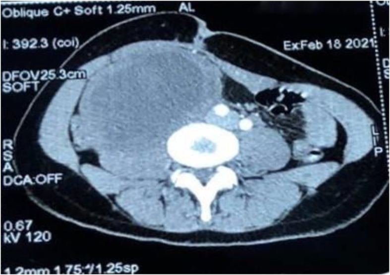
A 25-year-old woman presented to our department with a 2-year history of abdominal pain that was localized in the right flank without radiation, constipation/diarrhea or externalized digestive hemorrhage. On physical examination, we found a painless palpable mass in the right hypochondrium extending to the right iliac fossa, measuring approximately 10 cm. The MRI and CT scan showed the presence of a large intra-abdominal oval formation in the right para-umbilical region. It was well limited, measuring 11069mm with discrete irregular contours, thickened wall and heterogeneous content mostly fluid. They also showed the presence of a cystic formation in the right ovary measuring 8452mm and extending over 76mm. The procedure consisted of resection of the retroperitoneal solid cystic mass, right ovariectomy and drainage of the right parietal-colic gutter by Salem sump tube. A laparotomy with a median incision above and below the umbilicus was performed. After the resection, the specimens were sent for anatomopathological examination which concluded that the retroperitoneal mass was a schwannoma and the ovarian mass was a serous cystadenoma.
Retroperitoneal schwannomas are rare tumors and a pre-operative diagnosis is often difficult. The diagnosis is most often fortuitous and late, given the latency of the tumor's evolution, and the definitive diagnosis is based on histopathologic examination. Herein we presented a case of retroperitoneal schwannoma and studied the features of this phenomenon on the basis of the literature.
Retroperitoneal schwannomas are rare. The diagnosis is often late at the stage of a large tumor. Radiologic findings are usually nondiagnostic. The treatment of choice is complete surgical excision. Prognosis is good but because of the risk of recurrence and malignant transformation, further follow-up is necessary.
施万细胞瘤起源于周围神经鞘的施万细胞,很少发生于腹膜后(占所有施万细胞瘤的3%)。患者通常无症状或仅有非特异性症状,术前准确诊断困难。施万细胞瘤通常为良性,但很少发生恶变。在此,我们报告一例腹膜后施万细胞瘤病例并复习相关文献。
一名25岁女性因腹痛2年就诊于我科,腹痛局限于右下腹,无放射痛,无便秘/腹泻或消化道出血。体格检查发现右季肋部可触及一无痛性肿块,延伸至右髂窝,大小约10cm。MRI和CT扫描显示右脐旁区域有一巨大腹腔内椭圆形肿物。边界清晰,大小为11069mm,轮廓不规则,壁增厚,内容物不均质,大部分为液体。还显示右卵巢有一大小为8452mm、延伸超过76mm的囊性肿物。手术包括切除腹膜后实性囊性肿块、右侧卵巢切除术以及通过塞勒姆引流管引流右结肠旁沟。行脐上下正中切口的剖腹手术。切除后,标本送解剖病理学检查,结果显示腹膜后肿块为施万细胞瘤,卵巢肿块为浆液性囊腺瘤。
腹膜后施万细胞瘤是罕见肿瘤,术前诊断往往困难。由于肿瘤发展隐匿,诊断多为偶然且较晚,最终诊断基于组织病理学检查。在此我们报告一例腹膜后施万细胞瘤病例,并根据文献研究该现象的特征。
腹膜后施万细胞瘤罕见。诊断通常在肿瘤较大时较晚。影像学表现通常无诊断价值。治疗选择是完整手术切除。预后良好,但由于有复发和恶变风险