International School for Advanced Studies (SISSA/ISAS), 34136, Trieste, Italy.
Dipartimento di Medicina Sperimentale e Clinica, University of Florence, 50139, Florence, Italy.
Mol Brain. 2021 Oct 25;14(1):159. doi: 10.1186/s13041-021-00868-6.
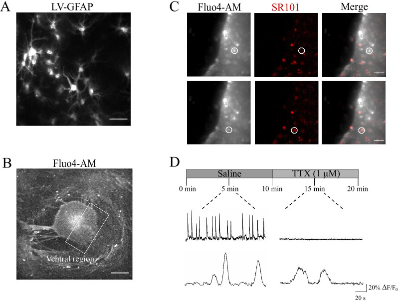
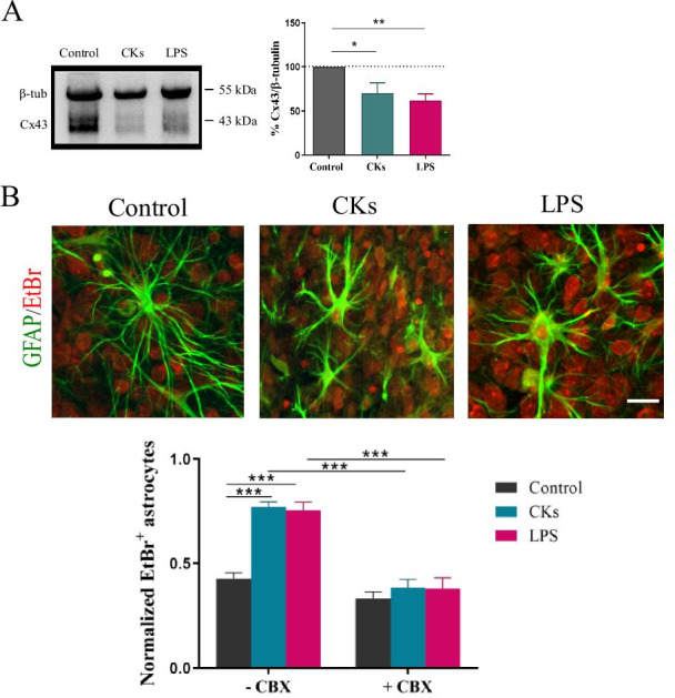
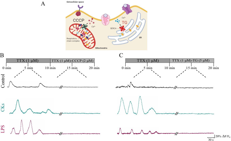
Neuroinflammation is an escalation factor shared by a vast range of central nervous system (CNS) pathologies, from neurodegenerative diseases to neuropsychiatric disorders. CNS immune status emerges by the integration of the responses of resident and not resident cells, leading to alterations in neural circuits functions. To explore spinal cord astrocyte reactivity to inflammatory threats we focused our study on the effects of local inflammation in a controlled micro-environment, the organotypic spinal slices, developed from the spinal cord of mouse embryos. These organ cultures represent a complex in vitro model where sensory-motor cytoarchitecture, synaptic properties and spinal cord resident cells, are retained in a 3D fashion and we recently exploit these cultures to model two diverse immune conditions in the CNS, involving different inflammatory networks and products. Here, we specifically focus on the tuning of calcium signaling in astrocytes by these diverse types of inflammation and we investigate the mechanisms which modulate intracellular calcium release and its spreading among astrocytes in the inflamed environment. Organotypic spinal cord slices are cultured for two or three weeks in vitro (WIV) and exposed for 6 h to a cocktail of cytokines (CKs), composed by tumor necrosis factor alpha (TNF-α), interleukin-1 beta (IL-1 β) and granulocyte macrophage-colony stimulating factor (GM-CSF), or to lipopolysaccharide (LPS). By live calcium imaging of the ventral horn, we document an increase in active astrocytes and in the occurrence of spontaneous calcium oscillations displayed by these cells when exposed to each inflammatory threat. Through several pharmacological treatments, we demonstrate that intracellular calcium sources and the activation of connexin 43 (Cx43) hemichannels have a pivotal role in increasing calcium intercellular communication in both CKs and LPS conditions, while the Cx43 gap junction communication is apparently reduced by the inflammatory treatments.
神经炎症是广泛的中枢神经系统(CNS)病理学的一个加剧因素,从神经退行性疾病到神经精神疾病。CNS 的免疫状态是通过常驻和非常驻细胞反应的整合而出现的,导致神经回路功能的改变。为了研究脊髓星形胶质细胞对炎症威胁的反应性,我们将研究重点放在局部炎症在受控微环境中的影响上,该微环境是从小鼠胚胎脊髓中开发的器官型脊髓切片。这些器官培养物代表了一种复杂的体外模型,其中感觉运动细胞结构、突触特性和脊髓常驻细胞以 3D 方式保留,我们最近利用这些培养物来模拟中枢神经系统中的两种不同的免疫状况,涉及不同的炎症网络和产物。在这里,我们特别关注这些不同类型的炎症对星形胶质细胞钙信号的调节,并研究调节细胞内钙释放及其在炎症环境中在星形胶质细胞之间传播的机制。器官型脊髓切片在体外培养 2 或 3 周(WIV),并暴露于细胞因子(CK)鸡尾酒中 6 小时,该鸡尾酒由肿瘤坏死因子-α(TNF-α)、白细胞介素-1β(IL-1β)和粒细胞巨噬细胞集落刺激因子(GM-CSF)组成,或脂多糖(LPS)。通过对腹角的活钙成像,我们记录到在暴露于每种炎症威胁时,活性星形胶质细胞增加,并且这些细胞显示自发钙振荡的发生。通过几种药理学处理,我们证明细胞内钙源和连接蛋白 43(Cx43)半通道的激活在 CK 和 LPS 条件下增加钙细胞间通讯中起关键作用,而 Cx43 间隙连接通讯显然被炎症处理所减少。