School of Basic Medicine, Tongji Medical College, Huazhong University of Science and Technology, Wuhan, 430030, P.R. China.
Department of Medical Laboratory, the Central Hospital of Wuhan, Tongji Medical College, Huazhong University of Science and Technology, Wuhan, 430014, P.R. China.
Commun Biol. 2021 Oct 25;4(1):1223. doi: 10.1038/s42003-021-02757-z.
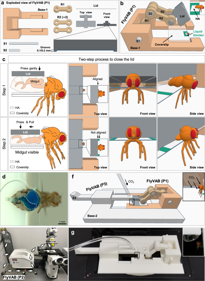
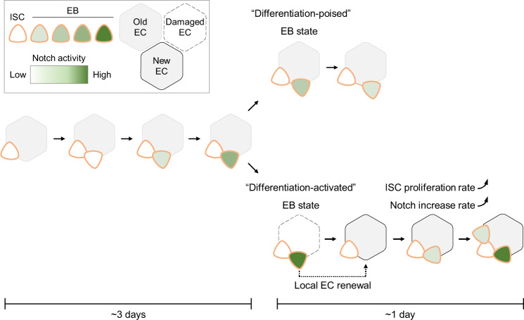
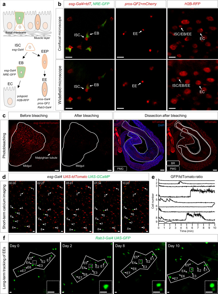
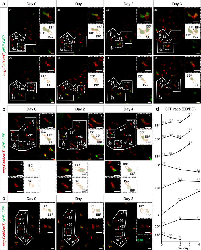
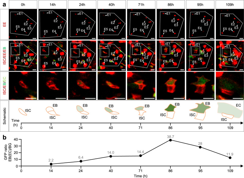
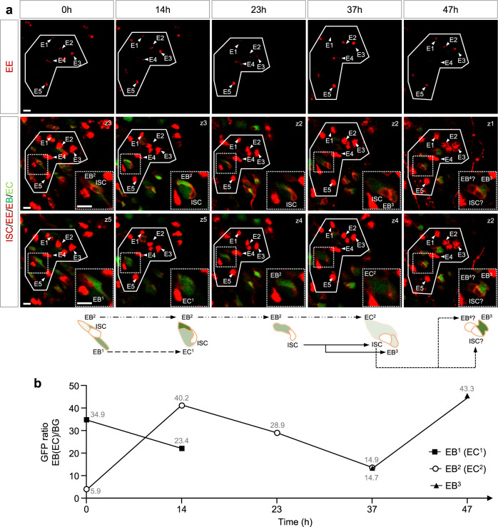
Aging or injury in Drosophila intestine promotes intestinal stem cell (ISC) proliferation and enteroblast (EB) differentiation. However, the manner the local physiology couples with dynamic EB differentiation assessed by traditional lineage tracing method is still vague. Therefore, we developed a 3D-printed platform "FlyVAB" for intravital imaging strategy that enables the visualization of the Drosophila posterior midgut at a single cell level across the ventral abdomen cuticle. Using ISCs in young and healthy midgut and enteroendocrine cells in age-associated hyperplastic midgut as reference coordinates, we traced ISC-EB-enterocyte lineages with Notch signaling reporter for multiple days. Our results reveal a "differentiation-poised" EB status correlated with slow ISC divisions and a "differentiation-activated" EB status correlated with ISC hyperplasia and rapid EB to enterocyte differentiation. Our FlyVAB imaging strategy opens the door to long-time intravital imaging of intestinal epithelium.
果蝇肠道的衰老或损伤会促进肠干细胞(ISC)的增殖和肠母细胞(EB)的分化。然而,通过传统的谱系追踪方法评估的局部生理学与动态 EB 分化之间的关联方式仍然不清楚。因此,我们开发了一种 3D 打印平台“FlyVAB”,用于活体成像策略,使我们能够在整个腹侧腹部表皮水平上以单细胞水平可视化果蝇后肠。使用年轻且健康的中肠中的 ISC 和与年龄相关的增生性中肠中的肠内分泌细胞作为参考坐标,我们使用 Notch 信号报告基因追踪了多个 ISC-EB-肠细胞谱系数日。我们的结果揭示了一种与 ISC 缓慢分裂相关的“分化准备”EB 状态,以及一种与 ISC 增生和 EB 向肠细胞分化快速相关的“分化激活”EB 状态。我们的 FlyVAB 成像策略为肠道上皮的长时间活体成像打开了大门。