Section of Pediatric Cardiology, Departments of Pediatrics and Physiology, New York Medical College, Valhalla, NY 10595, USA.
Med Sci (Basel). 2021 Sep 22;9(4):58. doi: 10.3390/medsci9040058.
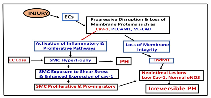
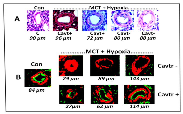
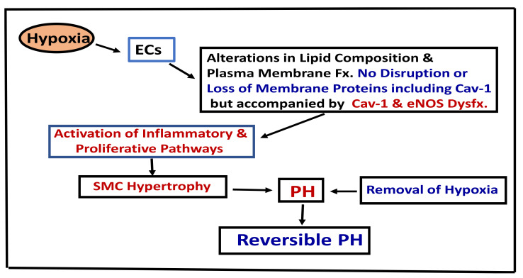
Pulmonary hypertension (PH) is a rare disease with a high morbidity and mortality rate. A number of systemic diseases and genetic mutations are known to lead to PH. The main features of PH are altered vascular relaxation responses and the activation of proliferative and anti-apoptotic pathways, resulting in pulmonary vascular remodeling, elevated pulmonary artery pressure, and right ventricular hypertrophy, ultimately leading to right heart failure and premature death. Important advances have been made in the field of pulmonary pathobiology, and several deregulated signaling pathways have been shown to be associated with PH. Clinical and experimental studies suggest that, irrespective of the underlying disease, endothelial cell disruption and/or dysfunction play a key role in the pathogenesis of PH. Endothelial caveolin-1, a cell membrane protein, interacts with and regulates several transcription factors and maintains homeostasis. Disruption of endothelial cells leads to the loss or dysfunction of endothelial caveolin-1, resulting in reciprocal activation of proliferative and inflammatory pathways, leading to cell proliferation, medial hypertrophy, and PH, which initiates PH and facilitates its progression. The disruption of endothelial cells, accompanied by the loss of endothelial caveolin-1, is accompanied by enhanced expression of caveolin-1 in smooth muscle cells (SMCs) that leads to pro-proliferative and pro-migratory responses, subsequently leading to neointima formation. The neointimal cells have low caveolin-1 and normal eNOS expression that may be responsible for promoting nitrosative and oxidative stress, furthering cell proliferation and metabolic alterations. These changes have been observed in human PH lungs and in experimental models of PH. In hypoxia-induced PH, there is no endothelial disruption, loss of endothelial caveolin-1, or enhanced expression of caveolin-1 in SMCs. Hypoxia induces alterations in membrane composition without caveolin-1 or any other membrane protein loss. However, caveolin-1 is dysfunctional, resulting in cell proliferation, medial hypertrophy, and PH. These alterations are reversible upon removal of hypoxia, provided there is no associated EC disruption. This review examined the role of caveolin-1 disruption and dysfunction in PH.
肺动脉高压(PH)是一种发病率和死亡率都很高的罕见疾病。已知许多系统性疾病和基因突变可导致 PH。PH 的主要特征是血管舒张反应改变和增殖及抗凋亡途径的激活,导致肺血管重构、肺动脉压升高和右心室肥厚,最终导致右心衰竭和过早死亡。在肺病理生物学领域取得了重要进展,已经证明几个失调的信号通路与 PH 有关。临床和实验研究表明,无论潜在疾病如何,内皮细胞破坏和/或功能障碍在 PH 的发病机制中起关键作用。内皮细胞 caveolin-1 是一种细胞膜蛋白,与几个转录因子相互作用并调节它们,维持着细胞内的稳态。内皮细胞的破坏导致内皮 caveolin-1 的丢失或功能障碍,从而导致增殖和炎症途径的反向激活,导致细胞增殖、中膜肥厚和 PH,启动 PH 并促进其进展。内皮细胞的破坏伴随着内皮 caveolin-1 的丢失,伴随着平滑肌细胞(SMC)中 caveolin-1 的表达增强,导致促增殖和促迁移反应,随后导致新内膜形成。新内膜细胞中 caveolin-1 表达水平较低,eNOS 表达正常,这可能是促进硝化和氧化应激、进一步促进细胞增殖和代谢改变的原因。这些变化在人类 PH 肺和 PH 实验模型中都有观察到。在缺氧诱导的 PH 中,没有内皮细胞的破坏、内皮 caveolin-1 的丢失或 SMC 中 caveolin-1 的表达增强。缺氧引起膜成分的改变,而没有 caveolin-1 或任何其他膜蛋白的丢失。然而,caveolin-1 功能失调,导致细胞增殖、中膜肥厚和 PH。在去除缺氧后,这些改变是可逆的,只要没有相关的 EC 破坏。本综述探讨了 caveolin-1 破坏和功能障碍在 PH 中的作用。