• 文献检索
  • 文档翻译
  • 深度研究
  • 学术资讯
  • Suppr Zotero 插件Zotero 插件
  • 邀请有礼
  • 套餐&价格
  • 历史记录
应用&插件
Suppr Zotero 插件Zotero 插件浏览器插件Mac 客户端Windows 客户端微信小程序
定价
高级版会员购买积分包购买API积分包
服务
文献检索文档翻译深度研究API 文档MCP 服务
关于我们
关于 Suppr公司介绍联系我们用户协议隐私条款
关注我们

Suppr 超能文献

核心技术专利:CN118964589B侵权必究
粤ICP备2023148730 号-1Suppr @ 2026

文献检索

告别复杂PubMed语法,用中文像聊天一样搜索,搜遍4000万医学文献。AI智能推荐,让科研检索更轻松。

立即免费搜索

文件翻译

保留排版,准确专业,支持PDF/Word/PPT等文件格式,支持 12+语言互译。

免费翻译文档

深度研究

AI帮你快速写综述,25分钟生成高质量综述,智能提取关键信息,辅助科研写作。

立即免费体验

圆锥角膜扩张作为圆锥角膜进展的预测指标。

Keratoconus enlargement as a predictor of keratoconus progression.

机构信息

Department of Ophthalmology, Centro Hospitalar Universitário São João, Porto, Portugal.

Department of Ophthalmology of São João Hospital, Avenida Prof. Hernâni Monteiro, 4202 - 451, Porto, Portugal.

出版信息

Sci Rep. 2021 Oct 26;11(1):21079. doi: 10.1038/s41598-021-00649-0.

DOI:10.1038/s41598-021-00649-0
PMID:34702891
原文链接:https://pmc.ncbi.nlm.nih.gov/articles/PMC8548552/
Abstract

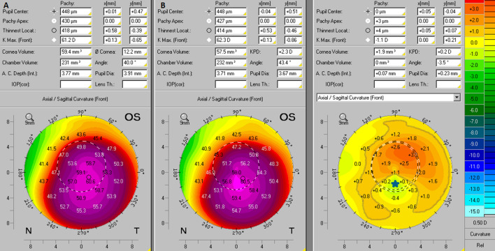
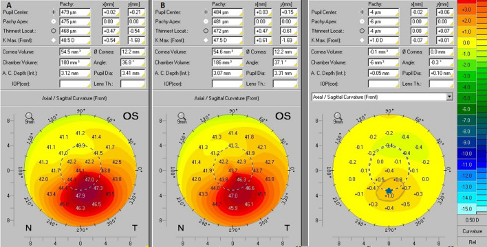
Numerous approaches have been designated to document progression in keratoconus, nevertheless there is no consistent or clear definition of ectasia progression. In this present study, we aim to evaluate Keratoconus Enlargement (KCE) as a parameter to document ectasia progression. We define KCE as an increase of more than 1D in the anterior curvature of non-apical corneal areas. We have designed a longitudinal study in 113 keratoconic eyes to assess keratoconus progression. KCE was compared with variables commonly used for detection of keratoconus progression like Kmax, Km, K2, PachyMin, D-Index, Corneal Astigmatism and PRC of 3.0 mm centered on the thinnest point. The variations of keratometric readings, D-index and ELEBmax showed positive associations with KCE. Evaluating the performance of Kmax, D-index and KCE as isolated parameters to document keratoconus progression we found a sensitivity of 49%, 82% and 77% and a specificity of 100%, 95% and 66% to detect keratoconus progression (p < 0.001 for all). This difference in sensitivity can be explained by the changes in keratoconus outside the small area represented by Kmax. The inclusion of KCE should be considered in the evaluation of keratoconus progression in conjunction with other variables to increase the reliability of our clinical evaluation.

摘要

已经有许多方法被指定用于记录圆锥角膜的进展,但对于扩张的进展还没有一致或明确的定义。在本研究中,我们旨在评估角膜扩张(KCE)作为记录扩张进展的参数。我们将 KCE 定义为非顶点角膜区域前曲率增加超过 1D。我们设计了一项针对 113 只圆锥角膜眼的纵向研究,以评估圆锥角膜的进展。将 KCE 与常用于检测圆锥角膜进展的变量进行比较,如 Kmax、Km、K2、PachyMin、D-Index、角膜散光和以最薄点为中心的 3.0mm 处的 PRC。角膜曲率读数、D-index 和 ELEBmax 的变化与 KCE 呈正相关。评估 Kmax、D-index 和 KCE 作为单独的参数来记录圆锥角膜的进展,我们发现它们的敏感性分别为 49%、82%和 77%,特异性分别为 100%、95%和 66%(所有 P 值均<0.001)。这种敏感性的差异可以用 Kmax 所代表的小区域以外的圆锥角膜变化来解释。在评估圆锥角膜进展时,应考虑将 KCE 纳入评估,同时结合其他变量,以提高我们临床评估的可靠性。

https://cdn.ncbi.nlm.nih.gov/pmc/blobs/2978/8548552/1eb768d9646b/41598_2021_649_Fig3_HTML.jpg
https://cdn.ncbi.nlm.nih.gov/pmc/blobs/2978/8548552/d41feb1a9eda/41598_2021_649_Fig1_HTML.jpg
https://cdn.ncbi.nlm.nih.gov/pmc/blobs/2978/8548552/8faa5f474bcf/41598_2021_649_Fig2_HTML.jpg
https://cdn.ncbi.nlm.nih.gov/pmc/blobs/2978/8548552/1eb768d9646b/41598_2021_649_Fig3_HTML.jpg
https://cdn.ncbi.nlm.nih.gov/pmc/blobs/2978/8548552/d41feb1a9eda/41598_2021_649_Fig1_HTML.jpg
https://cdn.ncbi.nlm.nih.gov/pmc/blobs/2978/8548552/8faa5f474bcf/41598_2021_649_Fig2_HTML.jpg
https://cdn.ncbi.nlm.nih.gov/pmc/blobs/2978/8548552/1eb768d9646b/41598_2021_649_Fig3_HTML.jpg

相似文献

1
Keratoconus enlargement as a predictor of keratoconus progression.圆锥角膜扩张作为圆锥角膜进展的预测指标。
Sci Rep. 2021 Oct 26;11(1):21079. doi: 10.1038/s41598-021-00649-0.
2
Comparison of eye-rubbing effect in keratoconic eyes and healthy eyes using Scheimpflug analysis and a dynamic bidirectional applanation device.应用 Scheimpflug 分析和动态双向眼压计比较圆锥角膜眼和正常眼的揉眼效应。
J Cataract Refract Surg. 2019 Aug;45(8):1156-1162. doi: 10.1016/j.jcrs.2019.03.017. Epub 2019 Jul 2.
3
Impact of classifying keratoconus location based on keratometry or pachymetry on progression parameters.基于角膜曲率或角膜厚度对圆锥角膜位置进行分类对进展参数的影响。
Clin Exp Optom. 2020 May;103(3):312-319. doi: 10.1111/cxo.12927. Epub 2019 Jun 11.
4
Keratometric indices for detecting the type of keratoconus: a combined discriminant analysis.用于检测圆锥角膜类型的角膜曲率指数:联合判别分析。
Clin Exp Optom. 2020 Jul;103(4):463-468. doi: 10.1111/cxo.13001. Epub 2019 Nov 26.
5
Characterization of posterior corneal astigmatism in a population with keratoconus.研究圆锥角膜患者后角膜散光的特征。
Semin Ophthalmol. 2020 Nov 16;35(7-8):352-357. doi: 10.1080/08820538.2020.1863436. Epub 2020 Dec 27.
6
The Outcome of Corneal Collagen Cross-Linking in Patients with Advanced Progressive Keratoconus: A 2-Year Follow-up Study.晚期进行性圆锥角膜患者角膜胶原交联的结果:一项为期2年的随访研究。
Middle East Afr J Ophthalmol. 2019 Jan-Mar;26(1):11-16. doi: 10.4103/meajo.MEAJO_101_18.
7
Comparison of variables measured with a Scheimpflug device for evaluation of progression and detection of keratoconus.应用 Scheimpflug 设备评估圆锥角膜进展和检测的变量比较。
Sci Rep. 2020 Nov 9;10(1):19308. doi: 10.1038/s41598-020-76020-6.
8
Longitudinal Evaluation of the Progression of Keratoconus Using a Novel Progression Display.使用新型进展显示评估圆锥角膜的进展:纵向研究。
Eye Contact Lens. 2019 Sep;45(5):324-330. doi: 10.1097/ICL.0000000000000582.
9
Longitudinal changes in corneal irregular astigmatism and visual acuity in eyes with keratoconus.圆锥角膜患者眼睛角膜不规则散光和视力的纵向变化
Jpn J Ophthalmol. 2007 Jul-Aug;51(4):265-9. doi: 10.1007/s10384-007-0453-2. Epub 2007 Aug 3.
10
[Characteristics of corneal topography in parents of keratoconus patients].[圆锥角膜患者父母的角膜地形图特征]
Zhonghua Yan Ke Za Zhi. 2020 Jun 11;56(6):456-464. doi: 10.3760/cma.j.cn112142-20191008-00200.

引用本文的文献

1
Progression and regression of keratoconus in an Iranian population.伊朗人群圆锥角膜的进展与消退
Sci Rep. 2025 Aug 5;15(1):28525. doi: 10.1038/s41598-025-08296-5.
2
Individual Risk Assessment and Prognostication of Outcomes After Corneal Cross-Linking.角膜交联术后个体风险评估及预后预测
J Ophthalmol. 2025 Jul 1;2025:3678453. doi: 10.1155/joph/3678453. eCollection 2025.
3
A novel metric to monitor early progression in keratoconus: epithelial backscatter.一种监测圆锥角膜早期进展的新指标:上皮后向散射。

本文引用的文献

1
Increased choroidal thickness is not a disease progression marker in keratoconus.脉络膜厚度增加不是圆锥角膜进展的标志物。
Sci Rep. 2020 Nov 17;10(1):19938. doi: 10.1038/s41598-020-77122-x.
2
Transepithelial Accelerated Corneal Collagen Cross-Linking: Two-Year Results.经上皮加速角膜胶原交联:两年结果
Clin Ophthalmol. 2020 Aug 14;14:2329-2337. doi: 10.2147/OPTH.S252940. eCollection 2020.
3
Increased Choroidal Thickness in Keratoconus Patients: Perspectives in the Disease Pathophysiology.圆锥角膜患者脉络膜厚度增加:疾病病理生理学视角
Int Ophthalmol. 2025 Jun 20;45(1):253. doi: 10.1007/s10792-025-03627-x.
4
Digital transformation of care for keratoconus patients: ML modeling structural outcomes of corneal collagen cross-linking.圆锥角膜患者护理的数字化转型:角膜胶原交联的机器学习建模结构结果
Front Med (Lausanne). 2025 Jun 4;12:1462653. doi: 10.3389/fmed.2025.1462653. eCollection 2025.
5
The Combined Utilization of Epithelial Thickness and Tomographic Parameters in Keratoconus Detection.角膜厚度与断层扫描参数联合应用于圆锥角膜检测
J Ophthalmol. 2025 Mar 18;2025:6647993. doi: 10.1155/joph/6647993. eCollection 2025.
6
Relationship between objective and subjective refraction measurements in patients with mild keratoconus.轻度圆锥角膜患者客观与主观验光测量结果之间的关系。
Int J Ophthalmol. 2025 Mar 18;18(3):398-403. doi: 10.18240/ijo.2025.03.04. eCollection 2025.
7
Strategic Management of Descemet's Membrane Perforation During DALK in Advanced Keratoconus.圆锥角膜晚期深层前板层角膜移植术中后弹力层穿孔的策略性管理
Rom J Ophthalmol. 2024 Oct-Dec;68(4):448-456. doi: 10.22336/rjo.2024.81.
8
Transepithelial Accelerated Crosslinking for Progressive Keratoconus: A Critical Analysis of Medium-Term Treatment Outcomes.用于进行性圆锥角膜的经上皮加速交联:中期治疗结果的批判性分析
Clin Ophthalmol. 2024 Feb 8;18:393-407. doi: 10.2147/OPTH.S450916. eCollection 2024.
9
Patterns in refractive error and treatment delay in keratoconus-An Australian study.圆锥角膜的屈光不正和治疗延迟模式:一项澳大利亚研究。
PLoS One. 2024 Jan 11;19(1):e0297268. doi: 10.1371/journal.pone.0297268. eCollection 2024.
10
Serum inflammatory biomarkers are associated with increased choroidal thickness in keratoconus.血清炎症生物标志物与圆锥角膜脉络膜厚度增加有关。
Sci Rep. 2023 Jul 5;13(1):10862. doi: 10.1038/s41598-023-37472-8.
J Ophthalmol. 2019 Dec 3;2019:2453931. doi: 10.1155/2019/2453931. eCollection 2019.
4
Transepithelial accelerated versus conventional corneal collagen crosslinking in patients with keratoconus: a comparative study.圆锥角膜患者经上皮加速与传统角膜胶原交联的比较研究
Clin Ophthalmol. 2019 Mar 1;13:445-452. doi: 10.2147/OPTH.S189183. eCollection 2019.
5
Involvement of anterior and posterior corneal surface area imbalance in the pathological change of keratoconus.角膜前表面和后表面区域失衡在圆锥角膜病变中的作用。
Sci Rep. 2018 Oct 9;8(1):14993. doi: 10.1038/s41598-018-33490-z.
6
Evaluation of keratoconus progression.评估圆锥角膜的进展。
Br J Ophthalmol. 2019 Apr;103(4):551-557. doi: 10.1136/bjophthalmol-2017-311651. Epub 2018 Jun 1.
7
Repeated Same-Day Versus Single Tomography Measurements of Keratoconic Eyes for Analysis of Disease Progression.用于分析圆锥角膜疾病进展的圆锥角膜眼同日重复测量与单次断层扫描测量对比研究
Cornea. 2018 Apr;37(4):474-479. doi: 10.1097/ICO.0000000000001513.
8
Identifying more reliable parameters for the detection of change during the follow-up of mild to moderate keratoconus patients.为轻度至中度圆锥角膜患者随访期间的病情变化检测确定更可靠的参数。
Eye Vis (Lond). 2017 Oct 25;4:24. doi: 10.1186/s40662-017-0089-3. eCollection 2017.
9
A review of keratoconus: Diagnosis, pathophysiology, and genetics.圆锥角膜的综述:诊断、病理生理学和遗传学。
Surv Ophthalmol. 2017 Nov-Dec;62(6):770-783. doi: 10.1016/j.survophthal.2017.06.009. Epub 2017 Jul 6.
10
Biomechanical Characterization of Subclinical Keratoconus Without Topographic or Tomographic Abnormalities.无地形图或断层扫描异常的亚临床圆锥角膜的生物力学特征
J Refract Surg. 2017 Jun 1;33(6):399-407. doi: 10.3928/1081597X-20170213-01.