Cui Ranliang, Wang Chaomin, Li Tiantian, Hua Jialei, Zhao Ting, Ren Li, Wang Yichao, Li Yueguo
Department of Clinical Laboratory, Tianjin Medical University Cancer Institute and Hospital, Tianjin's Clinical Research Center for Cancer, Key Laboratory of Cancer Prevention and Therapy, National Clinical Research Center for Cancer, Huanhuxi Road, Hexi District, Tianjin, 300060, China.
School of Medical Laboratory, Tianjin Medical University, Tianjin, China.
Cancer Cell Int. 2021 Oct 28;21(1):571. doi: 10.1186/s12935-021-02256-5.
The incidence and mortality of invasive breast cancer (IBC) are increasing annually. Hence, it is urgently needed to determine reliable biomarkers for not only monitoring curative effects, but evaluating prognosis. In present study, we aim to determine the potential role of Carboxypeptidase N1 (CPN1) in IBC tissues on chemotherapeutic efficacy and poor prognosis.
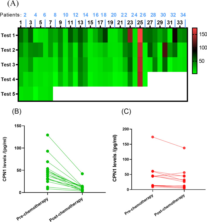
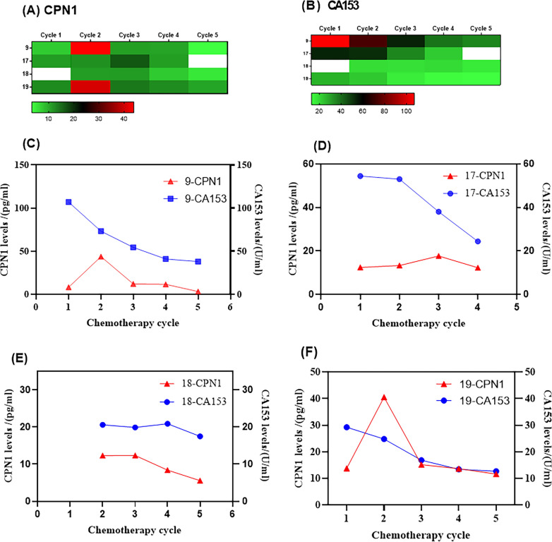
The expression level of CPN1 in IBC tissue samples (n = 123) was quantified by tissue microarray technique and immunohistochemical staining. Moreover, sera of IBC patients (n = 34) that underwent three to five consecutive chemotherapy sessions were collected. The patients were randomly stratified into a training (n = 15) as well as a validation group (n = 19). The expression of serum CA153 and CPN1 was quantified by electrochemiluminescence and ELISA assay, respectively.
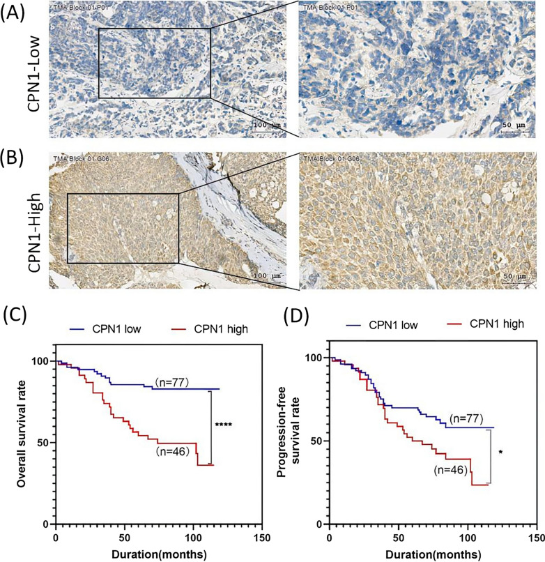
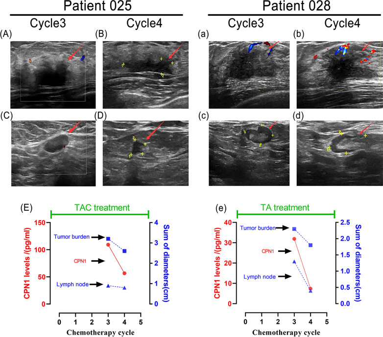
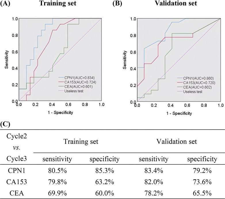
By univariate and multivariate Cox regression analysis, we show that CPN1 expression in IBC tissues, as an independent risk factor, is related to a poor overall survival (OS) and progression-free survival (PFS) (P < 0.05). Analysis of the data revealed that CPN1 over-expression could be consistently linked to adverse clinicopathological features such as lymph node metastasis and the pathological stage (pTNM) (P < 0.05). The serum CPN1 level trajectory of individual patients generally decreased during chemotherapy. In line with these findings were changes in the follow-up ultrasonography and a consistent decrease in serum CPN1 levels. The comparison of the area under the receiver operating curves (ROC) revealed that CPN1 has a better surveillance value than CA153 in the training (AUC = 0.834 vs. AUC= 0.724) as well as the validation set (AUC = 0.860 vs. AUC = 0.720) when comparing cycle2 versus cycle3.
CPN1 is a suitable potential biomarker for chemotherapeutic surveillance purposes as well as being an appropriate prognostic indicator which would support an improved chemotherapy regimen.
浸润性乳腺癌(IBC)的发病率和死亡率逐年上升。因此,迫切需要确定可靠的生物标志物,不仅用于监测疗效,还用于评估预后。在本研究中,我们旨在确定羧肽酶N1(CPN1)在IBC组织中对化疗疗效和不良预后的潜在作用。
采用组织芯片技术和免疫组化染色定量IBC组织样本(n = 123)中CPN1的表达水平。此外,收集了接受三到五个连续化疗疗程的IBC患者(n = 34)的血清。患者被随机分层为训练组(n = 15)和验证组(n = 19)。分别通过电化学发光和ELISA法对血清CA153和CPN1的表达进行定量。
通过单因素和多因素Cox回归分析,我们发现IBC组织中CPN1的表达作为独立危险因素,与较差的总生存期(OS)和无进展生存期(PFS)相关(P < 0.05)。数据分析显示,CPN1过表达可能与淋巴结转移和病理分期(pTNM)等不良临床病理特征一致相关(P < 0.05)。个体患者的血清CPN1水平轨迹在化疗期间通常下降。与这些发现一致的是随访超声检查的变化以及血清CPN1水平的持续下降。在比较第2周期和第3周期时,受试者操作曲线(ROC)下面积的比较显示,CPN1在训练组(AUC = 0.834 vs. AUC = 0.724)和验证组(AUC = 0.860 vs. AUC = 0.720)中比CA153具有更好的监测价值。
CPN1是用于化疗监测目的的合适潜在生物标志物,也是支持改进化疗方案的合适预后指标。