• 文献检索
  • 文档翻译
  • 深度研究
  • 学术资讯
  • Suppr Zotero 插件Zotero 插件
  • 邀请有礼
  • 套餐&价格
  • 历史记录
应用&插件
Suppr Zotero 插件Zotero 插件浏览器插件Mac 客户端Windows 客户端微信小程序
定价
高级版会员购买积分包购买API积分包
服务
文献检索文档翻译深度研究API 文档MCP 服务
关于我们
关于 Suppr公司介绍联系我们用户协议隐私条款
关注我们

Suppr 超能文献

核心技术专利:CN118964589B侵权必究
粤ICP备2023148730 号-1Suppr @ 2026

文献检索

告别复杂PubMed语法,用中文像聊天一样搜索,搜遍4000万医学文献。AI智能推荐,让科研检索更轻松。

立即免费搜索

文件翻译

保留排版,准确专业,支持PDF/Word/PPT等文件格式,支持 12+语言互译。

免费翻译文档

深度研究

AI帮你快速写综述,25分钟生成高质量综述,智能提取关键信息,辅助科研写作。

立即免费体验

瑞士-德国人群 101 颗下颌尖牙的微观 CT 内部形态:一项离体研究。

Internal morphology of 101 mandibular canines of a Swiss-German population by means of micro-CT: An ex vivo study.

机构信息

Department of Restorative, Preventive and Pediatric Dentistry, School of Dental Medicine, University of Bern, Freiburgstrasse 7, 3010, Bern, Switzerland.

Department of Periodontology and Operative Dentistry, University Medical Center of the Johannes Gutenberg-University Mainz, Mainz, Germany.

出版信息

Sci Rep. 2021 Oct 28;11(1):21281. doi: 10.1038/s41598-021-00758-w.

DOI:10.1038/s41598-021-00758-w
PMID:34711902
原文链接:https://pmc.ncbi.nlm.nih.gov/articles/PMC8553763/
Abstract

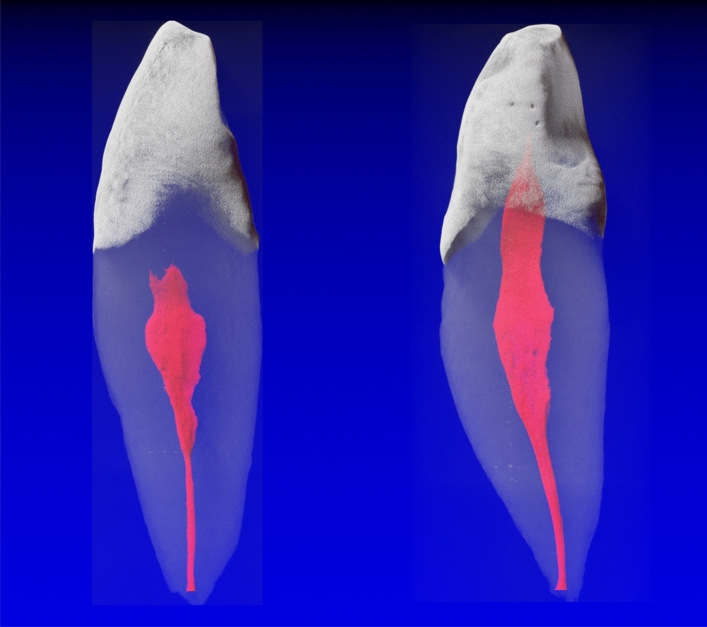
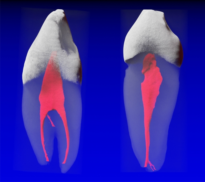
The aim of this study was to investigate the root canal system morphology by means of a root canal configuration (RCC) classification described with a four-digit system, the physiological foramen geometry and accessory canal frequency and morphology, of 101 mandibular canines (MaCa) of a Swiss-German population by means of micro-computed tomography. Micro-CT examination of the MaCa was performed and the obtained images analyzed with a 3D imaging software. In single-rooted MaCas, the most frequently observed RCCs were 1-1-1/1 (74.5%) and 1-1-1/2 (14.3%). Seven other RCCs were less frequently observed with a frequency from 4.1 to 1.0%. One physiological foramen was observed in 80.6% of the MaCas, two in 16.3%, three in 1.0% and four in 2.0%. Accessory and connecting canals were apparent only in the middle and apical root thirds. Two-rooted MaCas occurred less frequently (n = 3). When one physiological foramen was present, the mean size of the narrow and wide diameters were 0.28 mm (± 0.07) and 0.40 mm (± 0.11), while the distance between physiological and anatomical foramen was 0.45 mm (± 0.17). MaCas are predominantly single-rooted teeth with a 1-1-1/1 or 1-1-1/2 RCC. Most MaCas had one physiological foramen with an oval shape.

摘要

本研究旨在通过微计算机断层扫描(micro-computed tomography,micro-CT)研究瑞士-德国人群 101 颗下颌前磨牙(mandibular canines,MaCa)的根管形态、生理根尖孔形态和副根管频率及形态,采用四位数系统描述根管构型(root canal configuration,RCC)分类。对 MaCa 进行 micro-CT 检查,并用 3D 成像软件对获得的图像进行分析。在单根 MaCa 中,最常见的 RCC 为 1-1-1/1(74.5%)和 1-1-1/2(14.3%)。其他 7 种 RCC 的频率从 4.1%到 1.0%不等,相对较少见。80.6%的 MaCa 只有一个生理根尖孔,16.3%的 MaCa 有两个,1.0%的 MaCa 有三个,2.0%的 MaCa 有四个。副根管和连接根管仅在前磨牙的中、根尖三分之一处明显可见。双根 MaCa 较少见(n=3)。当存在一个生理根尖孔时,窄径和宽径的平均值分别为 0.28mm(±0.07)和 0.40mm(±0.11),而生理根尖孔和解剖根尖孔之间的距离为 0.45mm(±0.17)。MaCa 主要为具有 1-1-1/1 或 1-1-1/2 RCC 的单根牙齿。大多数 MaCa 只有一个呈椭圆形的生理根尖孔。

https://cdn.ncbi.nlm.nih.gov/pmc/blobs/338e/8553763/38c9255c67ef/41598_2021_758_Fig2_HTML.jpg
https://cdn.ncbi.nlm.nih.gov/pmc/blobs/338e/8553763/5d739729a0e2/41598_2021_758_Fig1_HTML.jpg
https://cdn.ncbi.nlm.nih.gov/pmc/blobs/338e/8553763/38c9255c67ef/41598_2021_758_Fig2_HTML.jpg
https://cdn.ncbi.nlm.nih.gov/pmc/blobs/338e/8553763/5d739729a0e2/41598_2021_758_Fig1_HTML.jpg
https://cdn.ncbi.nlm.nih.gov/pmc/blobs/338e/8553763/38c9255c67ef/41598_2021_758_Fig2_HTML.jpg

相似文献

1
Internal morphology of 101 mandibular canines of a Swiss-German population by means of micro-CT: An ex vivo study.瑞士-德国人群 101 颗下颌尖牙的微观 CT 内部形态:一项离体研究。
Sci Rep. 2021 Oct 28;11(1):21281. doi: 10.1038/s41598-021-00758-w.
2
3-dimensional Analysis and Literature Review of the Root Canal Morphology and Physiological Foramen Geometry of 125 Mandibular Incisors by Means of Micro-Computed Tomography in a German Population.德国人群 125 颗下颌切牙的根管形态和生理性根尖孔几何结构的三维分析和文献回顾:基于微计算机断层扫描技术。
J Endod. 2020 Feb;46(2):184-191. doi: 10.1016/j.joen.2019.11.006. Epub 2019 Dec 27.
3
3-Dimensional Analysis and Systematic Review of Root Canal Morphology and Physiological Foramen Geometry of 109 Mandibular First Premolars by Micro-computed Tomography in a Mixed Swiss-German Population.109 颗瑞士-德国混合人群下颌第一前磨牙的根管形态和生理性根尖孔几何结构的三维分析和系统评价。
J Endod. 2020 Jun;46(6):801-809. doi: 10.1016/j.joen.2020.03.002. Epub 2020 Apr 15.
4
Root Canal Morphology of 116 Maxillary Second Premolars by Micro-Computed Tomography in a Mixed Swiss-German Population with Systematic Review.瑞士-德国混合人群上颌第二前磨牙的根管形态的微计算机断层扫描研究及系统评价。
J Endod. 2020 Nov;46(11):1639-1647. doi: 10.1016/j.joen.2020.08.012. Epub 2020 Aug 18.
5
An Ex Vivo Study of Root Canal System Configuration and Morphology of 115 Maxillary First Premolars.115 颗上颌第一前磨牙根管系统形态的离体研究。
J Endod. 2020 Jun;46(6):794-800. doi: 10.1016/j.joen.2020.03.001. Epub 2020 Apr 13.
6
Root Canal Morphology and Configuration of 118 Mandibular First Molars by Means of Micro-Computed Tomography: An Ex Vivo Study.通过微计算机断层扫描对118颗下颌第一磨牙根管形态和结构的体外研究
J Endod. 2016 Apr;42(4):610-4. doi: 10.1016/j.joen.2016.01.004. Epub 2016 Feb 20.
7
Internal Morphology of Mandibular Second Premolars Using Micro-Computed Tomography.使用微型计算机断层扫描技术对下颌第二前磨牙的内部形态进行研究
J Imaging. 2023 Nov 23;9(12):257. doi: 10.3390/jimaging9120257.
8
Micro-computed tomographic analysis of the morphology of maxillary lateral incisors.上颌侧切牙形态的微计算机断层分析。
Clin Oral Investig. 2024 May 23;28(6):335. doi: 10.1007/s00784-024-05727-x.
9
Microcomputed tomography analysis of the root canal morphology of single-rooted mandibular canines.单根下颌尖牙根管形态的微计算机断层扫描分析
Int Endod J. 2013 Sep;46(9):800-7. doi: 10.1111/iej.12061. Epub 2013 Feb 12.
10
Micro-computed tomographic analysis of the root canal morphology of the distal root of mandibular first molar.下颌第一磨牙远中根根管形态的显微计算机断层扫描分析
J Endod. 2015 Feb;41(2):231-6. doi: 10.1016/j.joen.2014.09.024. Epub 2014 Nov 12.

引用本文的文献

1
Micro-computed tomographic analysis of the morphology of maxillary canines.上颌尖牙形态的显微计算机断层扫描分析
Sci Rep. 2025 Feb 3;15(1):4111. doi: 10.1038/s41598-024-84877-0.
2
Evaluation of root and canal morphologies of permanent canines in a Saudi Arabian sub population using cone-beam computed tomography.使用锥形束计算机断层扫描评估沙特阿拉伯亚人群中恒牙犬齿的根管形态。
J Dent Sci. 2022 Oct;17(4):1626-1633. doi: 10.1016/j.jds.2022.03.012. Epub 2022 Apr 9.
3
Root Morphology and Canal Configuration of Permanent Canines Among Saudi Population: Systematic Review and Comparison with Worldwide Studies.

本文引用的文献

1
Automated segmentation and description of the internal morphology of human permanent teeth by means of micro-CT.利用 micro-CT 对人恒磨牙内部形态进行自动分割和描述。
BMC Oral Health. 2021 Apr 12;21(1):185. doi: 10.1186/s12903-021-01551-x.
2
Morphometric analysis of the apical foramina in extracted human teeth.人牙根尖孔的形态计量分析。
Folia Morphol (Warsz). 2022;81(1):212-219. doi: 10.5603/FM.a2020.0143. Epub 2020 Dec 17.
3
Root and canal morphology of the anterior permanent dentition in Malaysian population using two classification systems: A CBCT clinical study.
沙特人群中恒牙犬齿的牙根形态和根管形态:系统评价及与全球研究的比较
Int J Gen Med. 2022 Aug 29;15:6849-6860. doi: 10.2147/IJGM.S380084. eCollection 2022.
4
Root and Root Canal Configuration Characterization Using Microcomputed Tomography: A Systematic Review.使用微型计算机断层扫描对牙根和根管形态特征进行表征:一项系统评价。
J Clin Med. 2022 Apr 20;11(9):2287. doi: 10.3390/jcm11092287.
马来西亚人群前永久恒牙的根和根管形态:两种分类系统的 CBCT 临床研究。
Aust Endod J. 2021 Aug;47(2):202-216. doi: 10.1111/aej.12454. Epub 2020 Nov 7.
4
Root canal morphology of mandibular canines in the Syrian population: A CBCT Assessment.叙利亚人群下颌尖牙的根管形态:CBCT评估
J Family Med Prim Care. 2020 Feb 28;9(2):552-555. doi: 10.4103/jfmpc.jfmpc_655_19. eCollection 2020 Feb.
5
Micro-computed tomographic assessment of the variability and morphological features of root canal system and their ramifications.根管系统及其分支的可变性和形态特征的微计算机断层评估。
J Appl Oral Sci. 2020 Feb 7;28:e20190393. doi: 10.1590/1678-7757-2019-0393. eCollection 2020.
6
3-dimensional Analysis and Literature Review of the Root Canal Morphology and Physiological Foramen Geometry of 125 Mandibular Incisors by Means of Micro-Computed Tomography in a German Population.德国人群 125 颗下颌切牙的根管形态和生理性根尖孔几何结构的三维分析和文献回顾:基于微计算机断层扫描技术。
J Endod. 2020 Feb;46(2):184-191. doi: 10.1016/j.joen.2019.11.006. Epub 2019 Dec 27.
7
Prevalence of a Second Root and Canal in Mandibular and Maxillary Canines in a Saudi Arabian Population: A Cone-beam Computed Tomography Study.沙特阿拉伯人群下颌和上颌尖牙第二牙根和根管的患病率:一项锥形束计算机断层扫描研究
J Contemp Dent Pract. 2019 Jul 1;20(7):773-777.
8
Ten simple rules for writing and sharing computational analyses in Jupyter Notebooks.在Jupyter Notebook中撰写和分享计算分析的十条简单规则。
PLoS Comput Biol. 2019 Jul 25;15(7):e1007007. doi: 10.1371/journal.pcbi.1007007. eCollection 2019 Jul.
9
The internal root canal morphology of single-rooted mandibular canines revealed by micro-computed tomography.微计算机断层扫描显示的单根下颌尖牙根管内部形态
J Conserv Dent. 2018 Nov-Dec;21(6):588-591. doi: 10.4103/JCD.JCD_313_18.
10
Mandibular first premolar apical morphology: A stereomicroscopic study.下颌第一前磨牙根尖形态:一项实体显微镜研究。
Aust Endod J. 2019 Aug;45(2):233-240. doi: 10.1111/aej.12313. Epub 2018 Nov 6.