Ma Shumei, Fu Xinxin, Liu Lin, Liu Yi, Feng Hao, Jiang Heya, Liu Xiaomei, Liu Rui, Liang Zhenzhen, Li Mengke, Tian Zhujun, Hu Boqi, Bai Yongheng, Liang Bing, Liu Xiaodong
School of Public Health and Management, Wenzhou Medical University, Wenzhou, China.
NHC Key Laboratory of Radiobiology, Jilin University, Changchun, China.
Front Cell Dev Biol. 2021 Oct 14;9:723801. doi: 10.3389/fcell.2021.723801. eCollection 2021.
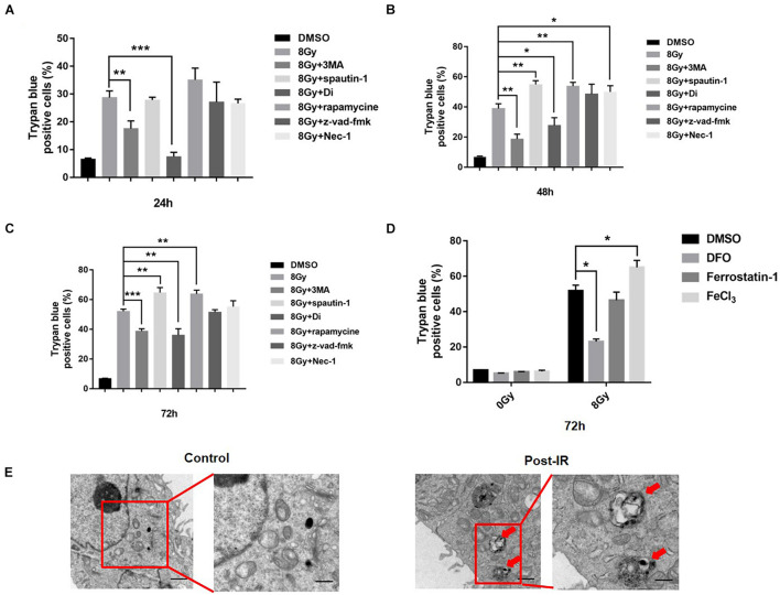
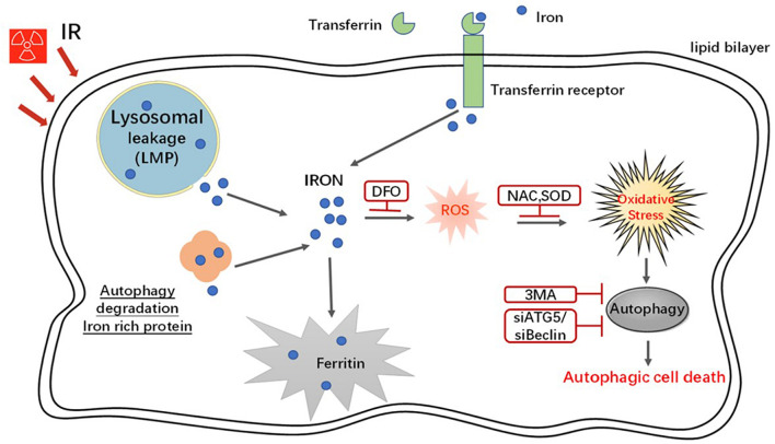
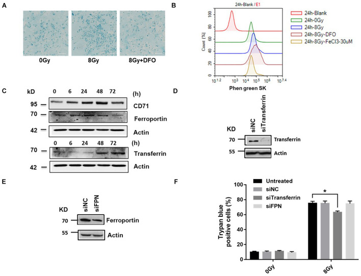
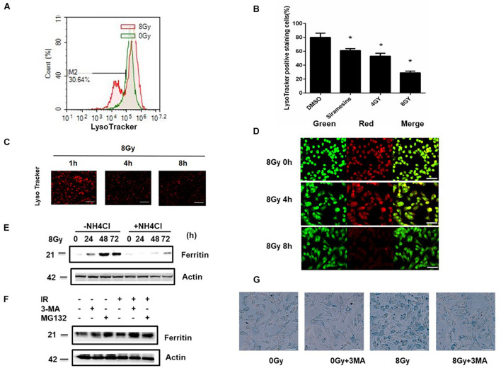
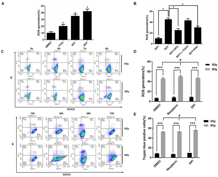
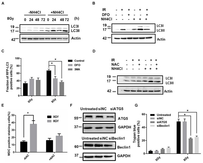
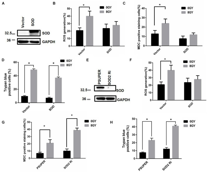
In radiation oncology, ionizing radiation is used to kill cancer cells, in other words, the induction of different types of cell death. To investigate this cellular death and the associated iron accumulation, the transfer, release, and participation of iron after radiation treatment was analyzed. We found that radiation-induced cell death varied in different breast cancer cells and autophagy was induced in MDA-MB-231 and BT549 cells (triple negative breast cancer cell line) rather than in MCF-7 and zr-75 cells. Iron chelator deferoxamine (DFO), the autophagy inhibitor 3MA, silencing of the autophagy-related genes ATG5, and Beclin 1 could decrease radiation induced cell death in MDA-MB-231 cells, while inhibitors of apoptosis such as Z-VAD-FMK, ferroptosis inhibitor ferrostatin-1 (Fer-1), and necroptosis inhibitor Necrostatin-1 showed no change. This suggests the occurrence of autophagic cell death. Furthermore, we found that iron accumulation and iron regulatory proteins, including transferrin (Tf), transferrin receptor (CD71), and Ferritin (FTH), increased after radiation treatment, and the silencing of transferrin decreased radiation-induced cell death. In addition, radiation increased lysosomal membrane permeabilization (LMP) and the release of lysosomal iron and cathepsins, while cathepsins silencing failed to change cell viability. Radiation-induced iron accumulation increased Reactive oxygen species (ROS) generation via the Fenton reaction and increased autophagy in a time-dependent manner. DFO, -acetylcysteine (NAC), and overexpression of superoxide dismutase 2 (SOD2) decreased ROS generation, autophagy, and cell death. To summarize, for the first time, we found that radiation-induced autophagic cell death was iron-dependent in breast cancer MDA-MB-231 cells. These results provide new insights into the cell death process of cancers and might conduce to the development and application of novel therapeutic strategies for patients with apoptosis-resistant breast cancer.
在放射肿瘤学中,电离辐射用于杀死癌细胞,也就是说,诱导不同类型的细胞死亡。为了研究这种细胞死亡以及相关的铁积累,分析了放射治疗后铁的转运、释放和参与情况。我们发现,辐射诱导的细胞死亡在不同的乳腺癌细胞中有所不同,自噬在MDA-MB-231和BT549细胞(三阴性乳腺癌细胞系)中被诱导,而在MCF-7和ZR-75细胞中未被诱导。铁螯合剂去铁胺(DFO)、自噬抑制剂3-甲基腺嘌呤(3MA)、自噬相关基因ATG5和Beclin 1的沉默可降低MDA-MB-231细胞中辐射诱导的细胞死亡,而凋亡抑制剂如Z-VAD-FMK、铁死亡抑制剂铁抑素-1(Fer-1)和坏死性凋亡抑制剂Necrostatin-1则无变化。这表明发生了自噬性细胞死亡。此外,我们发现放射治疗后铁积累以及包括转铁蛋白(Tf)、转铁蛋白受体(CD71)和铁蛋白(FTH)在内的铁调节蛋白增加,转铁蛋白的沉默降低了辐射诱导的细胞死亡。此外,辐射增加了溶酶体膜通透性(LMP)以及溶酶体铁和组织蛋白酶的释放,而组织蛋白酶的沉默未能改变细胞活力。辐射诱导的铁积累通过芬顿反应增加活性氧(ROS)的产生,并以时间依赖性方式增加自噬。DFO、N-乙酰半胱氨酸(NAC)和超氧化物歧化酶2(SOD2)的过表达降低了ROS的产生、自噬和细胞死亡。总之,我们首次发现,在乳腺癌MDA-MB-231细胞中,辐射诱导的自噬性细胞死亡是铁依赖性的。这些结果为癌症的细胞死亡过程提供了新的见解,并可能有助于开发和应用针对凋亡抗性乳腺癌患者的新型治疗策略。