Guys and St Thomas' NHS Foundation Trust, London, UK.
The Joint Department of Physics at the Institute of Cancer Research and the Royal Marsden NHS Foundation Trust, London, UK.
Br J Radiol. 2021 Dec;94(1128):20210350. doi: 10.1259/bjr.20210350. Epub 2021 Nov 2.
Quantify target volume delineation uncertainty for CT/MRI simulation and MRI-guided adaptive radiotherapy in rectal cancer. Define optimal imaging sequences for target delineation.
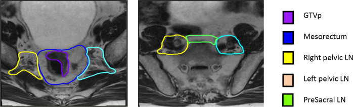
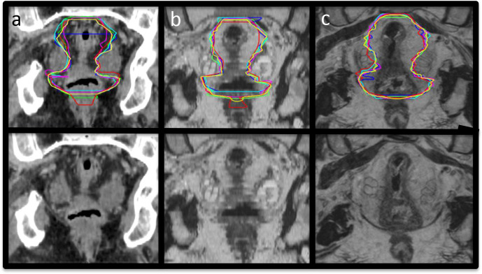
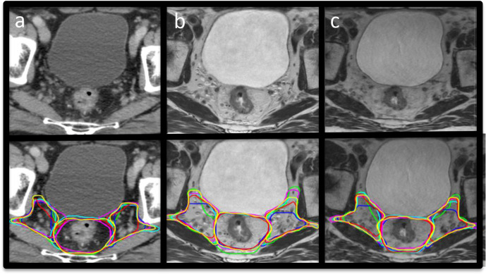
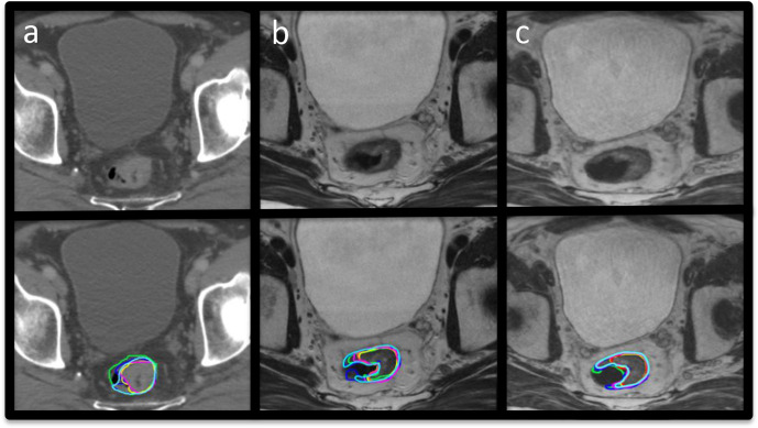
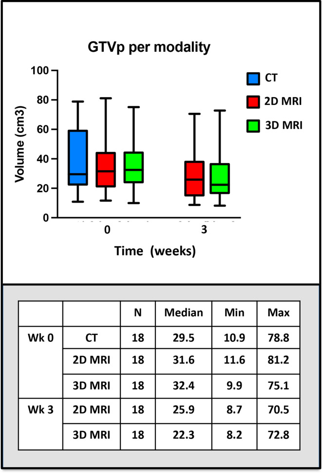
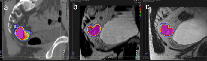
Six experienced radiation oncologists delineated clinical target volumes (CTVs) on CT and 2D and 3D-MRI in three patients with rectal cancer, using consensus contouring guidelines. Tumour GTV (GTVp) was also contoured on MRI acquired week 0 and 3 of radiotherapy. A STAPLE contour was created and volume and interobserver variability metrics were analysed.
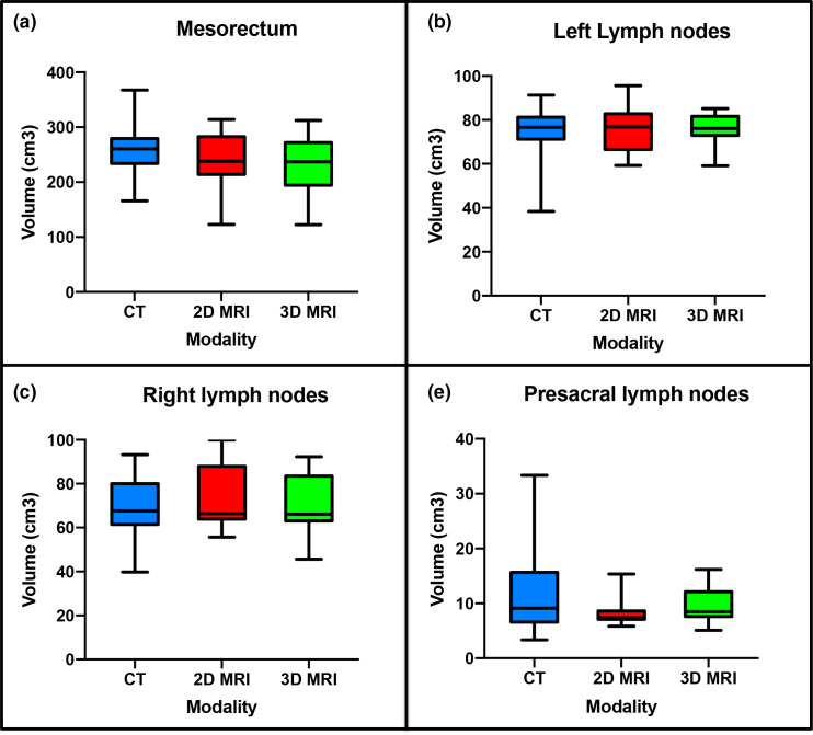
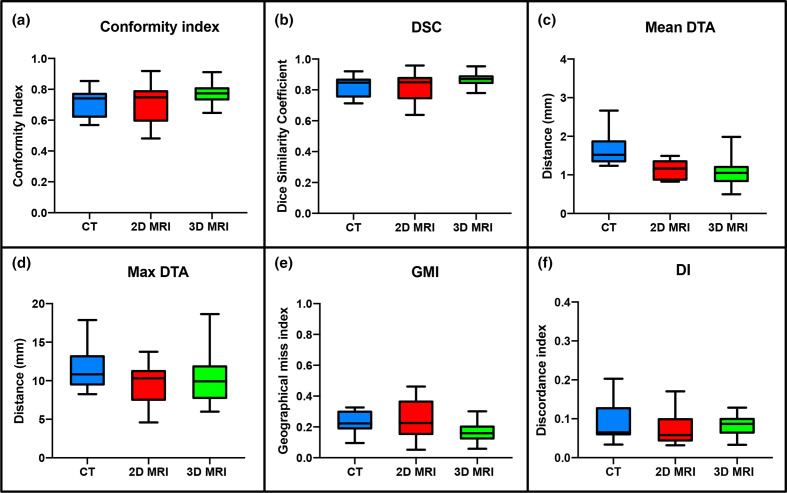
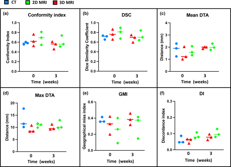
There were statistically significant differences in volume between observers for CT and 2D-MRI-defined CTVs ( < 0.05). There was no significant difference between observers on 3D-MRI. Significant differences in volume were seen between observers for both 2D and 3D-MRI-defined GTVp at weeks 0 and 3 ( < 0.05). Good interobserver agreement (IOA) was seen for CTVs delineated on all imaging modalities with best IOA on 3D-MRI; median Conformity index (CI) 0.74 for CT, 0.75 for 2D-MRI and 0.77 for 3D-MRI. IOA of MRI-defined GTVp week 0 was better compared to CT; CI 0.58 for CT, 0.62 for 2D-MRI and 0.7 for 3D-MRI. MRI-defined GTVp IOA week three was worse compared to week 0.
Delineation on MRI results in smaller volumes and better IOA week 0 compared to CT. 3D-MRI provides the best IOA in CTV and GTVp. MRI-defined GTVp on images acquired week 3 showed worse IOA compared to week 0. This highlights the need for consensus guidelines in GTVp delineation on MRI during treatment course in the context of dose escalation MRI-guided rectal boost studies.
Optimal MRI sequences for CT/MRI simulation and MRI-guided adaptive radiotherapy in rectal cancer have been defined.
量化 CT/MRI 模拟和 MRI 引导自适应放疗中直肠癌靶区勾画的不确定性。定义用于靶区勾画的最佳成像序列。
6 名经验丰富的放射肿瘤学家使用共识勾画指南在 3 名直肠癌患者的 CT 和 2D 及 3D-MRI 上勾画临床靶区(CTV)。还在放疗第 0 周和第 3 周勾画肿瘤 GTV(GTVp)。创建了一个 STAPLE 轮廓,并分析了体积和观察者间变异性指标。
在 CT 和 2D-MRI 定义的 CTV 上,观察者之间的体积存在统计学显著差异(<0.05)。在 3D-MRI 上,观察者之间没有显著差异。在第 0 周和第 3 周,观察者之间在 2D 和 3D-MRI 定义的 GTVp 体积上存在显著差异(<0.05)。在所有成像方式上勾画的 CTV 均具有良好的观察者间一致性(IOA),以 3D-MRI 最佳;中位适形指数(CI)分别为 CT 0.74、2D-MRI 0.75 和 3D-MRI 0.77。与 CT 相比,MRI 定义的 GTVp 第 0 周的 IOA 更好;CT 的 CI 为 0.58,2D-MRI 为 0.62,3D-MRI 为 0.7。MRI 定义的 GTVp 在第 3 周的 IOA 较第 0 周差。
与 CT 相比,MRI 勾画的体积更小,第 0 周的 IOA 更好。3D-MRI 为 CTV 和 GTVp 提供了最佳的 IOA。与第 0 周相比,第 3 周获取的图像上的 MRI 定义的 GTVp 的 IOA 更差。这凸显了在剂量递增 MRI 引导直肠增敏研究中,在治疗过程中需要在 MRI 上勾画 GTVp 共识指南的必要性。
已确定 CT/MRI 模拟和 MRI 引导自适应放疗中直肠癌的最佳 MRI 序列。