• 文献检索
  • 文档翻译
  • 深度研究
  • 学术资讯
  • Suppr Zotero 插件Zotero 插件
  • 邀请有礼
  • 套餐&价格
  • 历史记录
应用&插件
Suppr Zotero 插件Zotero 插件浏览器插件Mac 客户端Windows 客户端微信小程序
定价
高级版会员购买积分包购买API积分包
服务
文献检索文档翻译深度研究API 文档MCP 服务
关于我们
关于 Suppr公司介绍联系我们用户协议隐私条款
关注我们

Suppr 超能文献

核心技术专利:CN118964589B侵权必究
粤ICP备2023148730 号-1Suppr @ 2026

文献检索

告别复杂PubMed语法,用中文像聊天一样搜索,搜遍4000万医学文献。AI智能推荐,让科研检索更轻松。

立即免费搜索

文件翻译

保留排版,准确专业,支持PDF/Word/PPT等文件格式,支持 12+语言互译。

免费翻译文档

深度研究

AI帮你快速写综述,25分钟生成高质量综述,智能提取关键信息,辅助科研写作。

立即免费体验

新冠病毒病随访:康复三个月后胸部X光检查结果及其与临床和影像学的关系

COVID-19 follow-up: Chest X-ray findings with clinical and radiological relationship three months after recovery.

作者信息

Fogante M, Cavagna E, Rinaldi G

机构信息

Azienda Ospedaliera "Ospedali Riuniti", Ancona, Italy.

Ospedale "Infermi", Rimini, Italy.

出版信息

Radiography (Lond). 2022 May;28(2):531-536. doi: 10.1016/j.radi.2021.10.012. Epub 2021 Oct 22.

DOI:10.1016/j.radi.2021.10.012
PMID:34728138
原文链接:https://pmc.ncbi.nlm.nih.gov/articles/PMC8531194/
Abstract

INTRODUCTION

To evaluate the radiological sequelae of coronavirus disease (COVID-19) in a mid-term follow-up and investigate their relationship with clinical-radiological findings.

METHODS

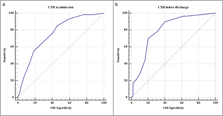
This prospective study included COVID-19 patients who underwent a CXR three months after discharge. The relationship between CXR score at three months after discharge and clinical findings and previous CXR scores, at admission and before the discharge, were evaluated. Then, based on mid-term follow-up CXR score, patients were divided in Group A (score = 0) and Group B (score≥1), and clinical-radiological findings were compared between two Groups. Finally, we calculated the CXR scores at admission and before the discharge with the highest sensitivity and specificity to predict normal and abnormal CXR score at mid-term follow-up.

RESULTS

The study included 119 patients, mean age 65.9 ± 14.6 years. The oxygen saturation (Sa) (p = 0.0006), the days of hospitalization (p < 0.0001) and the CXR score before the discharge (p = 0.0091) were independent factors to predict the mid-term follow-up CXR score. The Group A, 59 (49.6%) patients, had CXR scores at admission and before the discharge lower than Group B. The CXR scores at admission and before the discharge with the highest sensitivity and specificity to predict normal and abnormal CXR score at mid-term follow-up were, respectively, 3 and 2 (p < 0.0001).

CONCLUSIONS

The radiological abnormalities were present in about half patients three months after discharge, which had higher age, previous CXR scores and longer hospitalization. The S, days of hospitalization and previous CXR scores were independent factors for predicting the CXR at three months.

IMPLICATIONS FOR PRACTICE

The radiologist with CXR could play a central role in mid to long-term follow-up of COVID-19, assessing the radiological sequelae of patients and identifying those who might require a closer follow-up.

摘要

引言

在中期随访中评估冠状病毒病(COVID-19)的放射学后遗症,并研究它们与临床放射学表现之间的关系。

方法

这项前瞻性研究纳入了出院三个月后接受胸部X线检查(CXR)的COVID-19患者。评估出院三个月后的CXR评分与临床结果以及入院时和出院前的既往CXR评分之间的关系。然后,根据中期随访CXR评分,将患者分为A组(评分=0)和B组(评分≥1),并比较两组之间的临床放射学表现。最后,我们计算入院时和出院前具有最高敏感性和特异性的CXR评分,以预测中期随访时正常和异常的CXR评分。

结果

该研究纳入了119例患者,平均年龄65.9±14.6岁。血氧饱和度(Sa)(p=0.0006)、住院天数(p<0.0001)和出院前的CXR评分(p=0.0091)是预测中期随访CXR评分的独立因素。A组有59例(49.6%)患者,其入院时和出院前的CXR评分低于B组。在预测中期随访时正常和异常CXR评分方面,入院时和出院前具有最高敏感性和特异性的CXR评分分别为3分和2分(p<0.0001)。

结论

出院三个月后,约一半患者存在放射学异常,这些患者年龄较大、既往CXR评分较高且住院时间较长。Sa、住院天数和既往CXR评分是预测三个月时CXR的独立因素。

对实践的启示

进行CXR检查的放射科医生在COVID-19的中长期随访中可发挥核心作用,评估患者的放射学后遗症,并识别那些可能需要密切随访的患者。

https://cdn.ncbi.nlm.nih.gov/pmc/blobs/1296/8531194/20c522d85628/gr3_lrg.jpg
https://cdn.ncbi.nlm.nih.gov/pmc/blobs/1296/8531194/2b036da26a46/gr1_lrg.jpg
https://cdn.ncbi.nlm.nih.gov/pmc/blobs/1296/8531194/6e875e1284c6/gr2_lrg.jpg
https://cdn.ncbi.nlm.nih.gov/pmc/blobs/1296/8531194/20c522d85628/gr3_lrg.jpg
https://cdn.ncbi.nlm.nih.gov/pmc/blobs/1296/8531194/2b036da26a46/gr1_lrg.jpg
https://cdn.ncbi.nlm.nih.gov/pmc/blobs/1296/8531194/6e875e1284c6/gr2_lrg.jpg
https://cdn.ncbi.nlm.nih.gov/pmc/blobs/1296/8531194/20c522d85628/gr3_lrg.jpg

相似文献

1
COVID-19 follow-up: Chest X-ray findings with clinical and radiological relationship three months after recovery.新冠病毒病随访:康复三个月后胸部X光检查结果及其与临床和影像学的关系
Radiography (Lond). 2022 May;28(2):531-536. doi: 10.1016/j.radi.2021.10.012. Epub 2021 Oct 22.
2
Performance of a Severity Score on Admission Chest Radiography in Predicting Clinical Outcomes in Hospitalized Patients With Coronavirus Disease (COVID-19).入院胸部X光片严重程度评分对冠状病毒病(COVID-19)住院患者临床结局的预测作用
AJR Am J Roentgenol. 2021 Sep;217(3):623-632. doi: 10.2214/AJR.20.24801. Epub 2020 Oct 28.
3
Predicting the risk of chest radiograph abnormality 12-weeks post hospitalisation with SARS CoV-2 PCR confirmed COVID-19.预测 SARS CoV-2 PCR 确诊 COVID-19 住院后 12 周胸部 X 线异常的风险。
Respir Res. 2022 Oct 31;23(1):297. doi: 10.1186/s12931-022-02217-0.
4
COVID-2019 Pneumonia: Severity and distribution of lung changes observed on the initial chest X-ray as an indicator of final outcomes.COVID-19 肺炎:初始胸部 X 光片上观察到的肺部变化的严重程度和分布情况作为最终结果的指标。
Sultan Qaboos Univ Med J. 2022 Feb;22(1):98-105. doi: 10.18295/squmj.4.2021.061. Epub 2022 Feb 28.
5
Proposed Scoring System for Evaluating Clinico-radiological Severity of COVID- 19 using Plain Chest X- ray (CXR) changes (CO X-RADS): Preliminary results.基于胸部平片(CXR)改变的 COVID-19 临床-放射学严重程度评估用评分系统(CO X-RADS):初步结果。
Acta Biomed. 2020 Nov 10;91(4):e2020172. doi: 10.23750/abm.v91i4.10664.
6
Sextus chest radiograph severity score correlates to clinical outcomes in patients with COVID-19: A cross-sectional study.塞克斯图斯胸部 X 光片严重程度评分与 COVID-19 患者的临床结局相关:一项横断面研究。
Medicine (Baltimore). 2021 Nov 12;100(45):e27663. doi: 10.1097/MD.0000000000027663.
7
Observational study of clinico-radiological follow-up of COVID-19 pneumonia: a district general hospital experience in the UK.观察性研究:英国地区综合医院对 COVID-19 肺炎的临床-放射学随访经验。
BMC Infect Dis. 2021 Dec 8;21(1):1233. doi: 10.1186/s12879-021-06941-8.
8
Risk factors for persistent abnormality on chest radiographs at 12-weeks post hospitalisation with PCR confirmed COVID-19.PCR确诊的COVID-19患者住院12周后胸部X光片持续异常的危险因素。
Respir Res. 2021 May 21;22(1):157. doi: 10.1186/s12931-021-01750-8.
9
Which role for chest x-ray score in predicting the outcome in COVID-19 pneumonia?胸部X光评分在预测COVID-19肺炎预后方面起何种作用?
Eur Radiol. 2021 Jun;31(6):4016-4022. doi: 10.1007/s00330-020-07504-2. Epub 2020 Dec 2.
10
Association between chest X-ray score and clinical outcome in COVID-19 patients: A study on modified radiographic assessment of lung edema score (mRALE) in Indonesia.胸部 X 射线评分与 COVID-19 患者临床结局的关系:印度尼西亚改良放射性肺水肿评分(mRALE)的研究。
Narra J. 2024 Apr;4(1):e691. doi: 10.52225/narra.v4i1.691. Epub 2024 Apr 6.

引用本文的文献

1
Systolic Pulmonary Artery Pressure as Long-Term Mortality Predictor in Elderly Critically Ill with Severe COVID-19 Pneumonia.收缩期肺动脉压作为老年重症新型冠状病毒肺炎患者长期死亡率的预测指标
Viruses. 2025 Feb 11;17(2):244. doi: 10.3390/v17020244.
2
Clinico-radiological and pulmonary function assessment of post-COVID-19 patients with respiratory symptoms.有呼吸道症状的新冠后患者的临床影像学和肺功能评估
J Family Med Prim Care. 2024 Aug;13(8):2912-2920. doi: 10.4103/jfmpc.jfmpc_1721_23. Epub 2024 Jul 26.
3
Prevalence and patterns of post-COVID-19 symptoms in recovered patients of Delhi, India: a population-based study.

本文引用的文献

1
Features of severe COVID-19: A systematic review and meta-analysis.严重 COVID-19 的特征:系统评价和荟萃分析。
Eur J Clin Invest. 2020 Oct;50(10):e13378. doi: 10.1111/eci.13378. Epub 2020 Aug 29.
2
2019 novel coronavirus (COVID-19) pneumonia: CT manifestations and pattern of evolution in 110 patients in Jiangxi, China.2019 新型冠状病毒(COVID-19)肺炎:中国江西 110 例患者的 CT 表现和演变模式。
Eur Radiol. 2021 Feb;31(2):1059-1068. doi: 10.1007/s00330-020-07201-0. Epub 2020 Aug 27.
3
COVID-19 (Coronavirus Disease 2019): A New Coronavirus Disease.
印度德里康复患者中新冠后症状的患病率及模式:一项基于人群的研究
Osong Public Health Res Perspect. 2024 Jun;15(3):229-237. doi: 10.24171/j.phrp.2023.0251. Epub 2024 May 17.
4
[Lung Involvement Following Acute SARS-CoV-2 Infection].[新型严重急性呼吸综合征冠状病毒2型(SARS-CoV-2)急性感染后的肺部受累情况]
Open Respir Arch. 2022 Feb 8;4(2):100167. doi: 10.1016/j.opresp.2022.100167. eCollection 2022 Apr-Jun.
5
Health-related quality of life and radiological and functional lung changes of patients with COVID-19 Pneumonia 3 and 10 months after discharge.COVID-19 肺炎患者出院后 3 个月和 10 个月时的健康相关生活质量和肺部影像学及功能变化。
BMC Pulm Med. 2023 Jun 27;23(1):231. doi: 10.1186/s12890-023-02520-6.
6
The Short- and Long-Term Clinical, Radiological and Functional Consequences of COVID-19.《COVID-19 的短期和长期临床、影像学和功能后果》。
Arch Bronconeumol. 2022 Apr;58:32-38. doi: 10.1016/j.arbres.2022.03.006. Epub 2022 Apr 13.
新型冠状病毒肺炎(2019冠状病毒病):一种新型冠状病毒疾病
Infect Drug Resist. 2020 Aug 12;13:2819-2828. doi: 10.2147/IDR.S259279. eCollection 2020.
4
Respiratory follow-up of patients with COVID-19 pneumonia.COVID-19 肺炎患者的呼吸随访。
Thorax. 2020 Nov;75(11):1009-1016. doi: 10.1136/thoraxjnl-2020-215314. Epub 2020 Aug 24.
5
Follow-up study of the pulmonary function and related physiological characteristics of COVID-19 survivors three months after recovery.新冠康复者康复三个月后肺功能及相关生理特征的随访研究
EClinicalMedicine. 2020 Aug;25:100463. doi: 10.1016/j.eclinm.2020.100463. Epub 2020 Jul 15.
6
COVID-19 severity scoring systems in radiological imaging - a review.放射影像学中COVID-19严重程度评分系统——综述
Pol J Radiol. 2020 Jul 17;85:e361-e368. doi: 10.5114/pjr.2020.98009. eCollection 2020.
7
Severe acute respiratory syndrome coronavirus-2 (SARS-CoV-2) and coronavirus disease 19 (COVID-19) - anatomic pathology perspective on current knowledge.严重急性呼吸综合征冠状病毒 2 型(SARS-CoV-2)和 2019 冠状病毒病(COVID-19)——当前知识的解剖病理学视角。
Diagn Pathol. 2020 Aug 14;15(1):103. doi: 10.1186/s13000-020-01017-8.
8
Low physical functioning and impaired performance of activities of daily life in COVID-19 patients who survived hospitalisation.COVID-19 患者出院后身体机能下降,日常生活活动能力受损。
Eur Respir J. 2020 Oct 15;56(4). doi: 10.1183/13993003.02096-2020. Print 2020 Oct.
9
Dynamic changes of Chest CT follow-up in Coronavirus Disease-19 (COVID-19) pneumonia: relationship to clinical typing.新冠肺炎(COVID-19)肺炎的胸部 CT 随访动态变化:与临床分型的关系。
BMC Med Imaging. 2020 Aug 5;20(1):92. doi: 10.1186/s12880-020-00491-2.
10
Functional characteristics of patients with SARS-CoV-2 pneumonia at 30 days post-infection.感染 SARS-CoV-2 后 30 天肺炎患者的功能特征。
Eur Respir J. 2020 Aug 6;56(2). doi: 10.1183/13993003.01754-2020. Print 2020 Aug.