Monti Caterina Beatrice, Schiaffino Simone, Galimberti Ortiz Maria Del Mar, Capra Davide, Zanardo Moreno, De Benedictis Elena, Luporini Alberto Gianluigi, Spagnolo Pietro, Secchi Francesco, Sardanelli Francesco
Department of Biomedical Sciences for Health, Università Degli Studi Di Milano, Via Mangiagalli 31, 20133, Milano, Italy.
Unit of Radiology, IRCCS Policlinico San Donato, Via Morandi 30, 20097, San Donato Milanese, Italy.
Insights Imaging. 2021 Nov 6;12(1):161. doi: 10.1186/s13244-021-01069-4.
We investigated the radiodensity of epicardial (EAT), subcutaneous (SAT), and visceral adipose tissue (VAT) before and after treatment with anthracyclines in a population of breast cancer (BC) patients, and in controls not treated with anthracyclines, to detect a potential role of EAT density as a biomarker of changes related to chemotherapy cardiotoxicity.
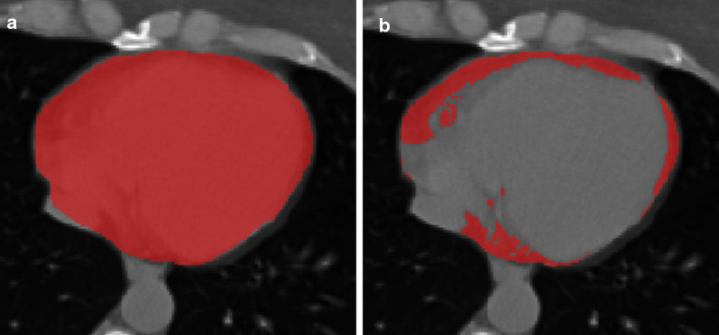
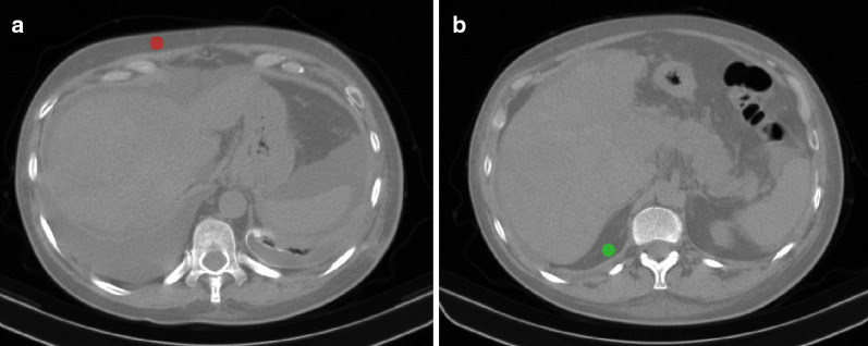
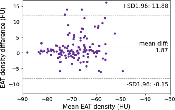
We reviewed BC patients treated with anthracyclines who underwent CT before (CT-t) and after (CT-t) chemotherapy, and age- and sex-matched controls who underwent two CT examinations at comparable intervals. On non-contrast scans, EAT was segmented contouring the pericardium and thresholding between -190 and -30 Hounsfield units (HU), and SAT and VAT were segmented with two 15-mm diameter regions of interest thresholded between -195 and -45 HU.
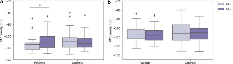
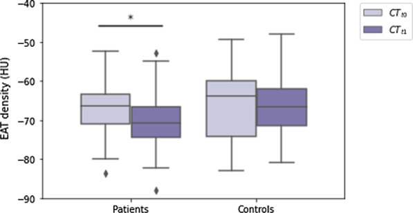
Thirty-two female patients and 32 controls were included. There were no differences in age (p = 0.439) and follow-up duration (p = 0.162) between patients and controls. Between CT-t and CT-t, EAT density decreased in BC patients (-66 HU, interquartile range [IQR] -71 to -63 HU, to -71 HU, IQR -75 to -66 HU, p = 0.003), while it did not vary in controls (p = 0.955). SAT density increased from CT-t to CT-t in BC patients (-107 HU, IQR -111 to -105 HU, to -105 HU, IQR -110 to -100 HU, p = 0.014), whereas it did not change in controls (p = 0.477). VAT density did not vary in either BC patients (p = 0.911) or controls (p = 0.627).
EAT density appears to be influenced by anthracycline treatment for BC, well known for its cardiotoxicity, shifting towards lower values indicative of a less active metabolism.
我们在一组乳腺癌(BC)患者以及未接受蒽环类药物治疗的对照组中,研究了蒽环类药物治疗前后心外膜脂肪组织(EAT)、皮下脂肪组织(SAT)和内脏脂肪组织(VAT)的放射密度,以检测EAT密度作为与化疗心脏毒性相关变化的生物标志物的潜在作用。
我们回顾了接受蒽环类药物治疗的BC患者,这些患者在化疗前(CT-t)和化疗后(CT-t)接受了CT检查,以及年龄和性别匹配的对照组,他们在可比的间隔时间内接受了两次CT检查。在非增强扫描中,通过勾勒心包轮廓并在-190至-30亨氏单位(HU)之间设置阈值来分割EAT,通过在两个直径为15毫米的感兴趣区域在-195至-45 HU之间设置阈值来分割SAT和VAT。
纳入了32名女性患者和32名对照组。患者和对照组之间在年龄(p = 0.439)和随访时间(p = 0.162)上没有差异。在CT-t和CT-t之间,BC患者的EAT密度降低(-66 HU,四分位间距[IQR]-71至-63 HU,降至-71 HU,IQR -75至-66 HU,p = 0.003)而对照组中其没有变化(p = 0.955)。BC患者的SAT密度从CT-t到CT-t增加(-107 HU,IQR -111至-105 HU,升至-105 HU,IQR -110至-100 HU,p = 0.014),而对照组中其没有变化(p = 0.477)。BC患者和对照组的VAT密度均没有变化(分别为p = 0.911和p = 0.627)。
EAT密度似乎受到以心脏毒性闻名的BC蒽环类药物治疗的影响,向更低值转变,表明代谢活性降低。