Brodtmann Amy, Werden Emilio, Khlif Mohamed Salah, Bird Laura J, Egorova Natalia, Veldsman Michele, Pardoe Heath, Jackson Graeme, Bradshaw Jennifer, Darby David, Cumming Toby, Churilov Leonid, Donnan Geoffrey
The Florey Institute of Neuroscience and Mental Health, University of Melbourne, Melbourne, VIC, Australia.
Melbourne Dementia Research Centre, Florey Institute and University of Melbourne, Parkville, VIC, Australia.
Front Neurol. 2021 Oct 22;12:754204. doi: 10.3389/fneur.2021.754204. eCollection 2021.
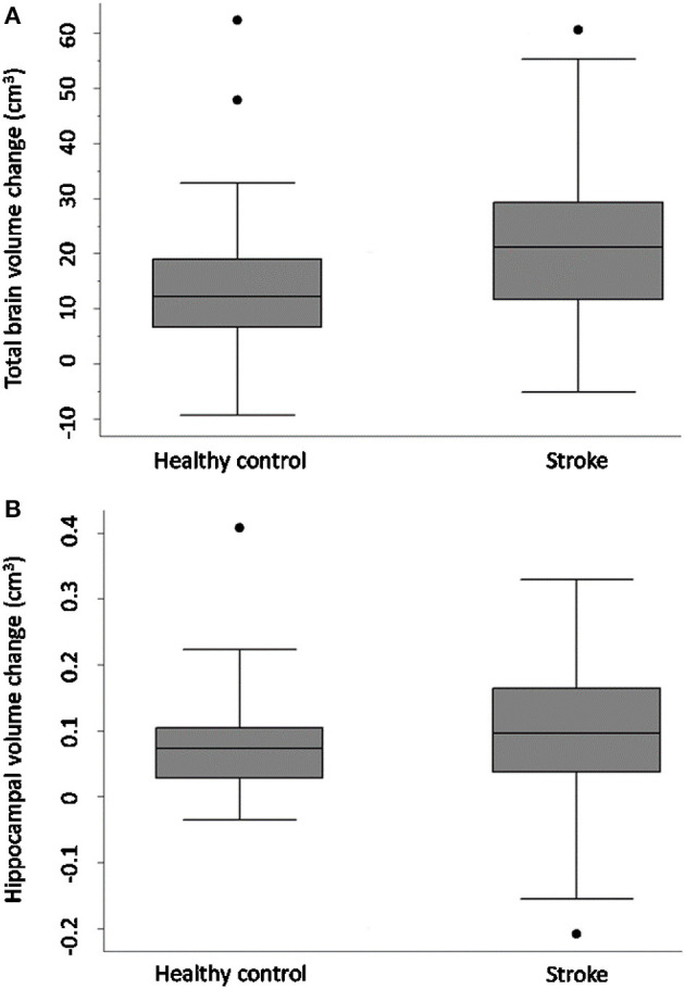
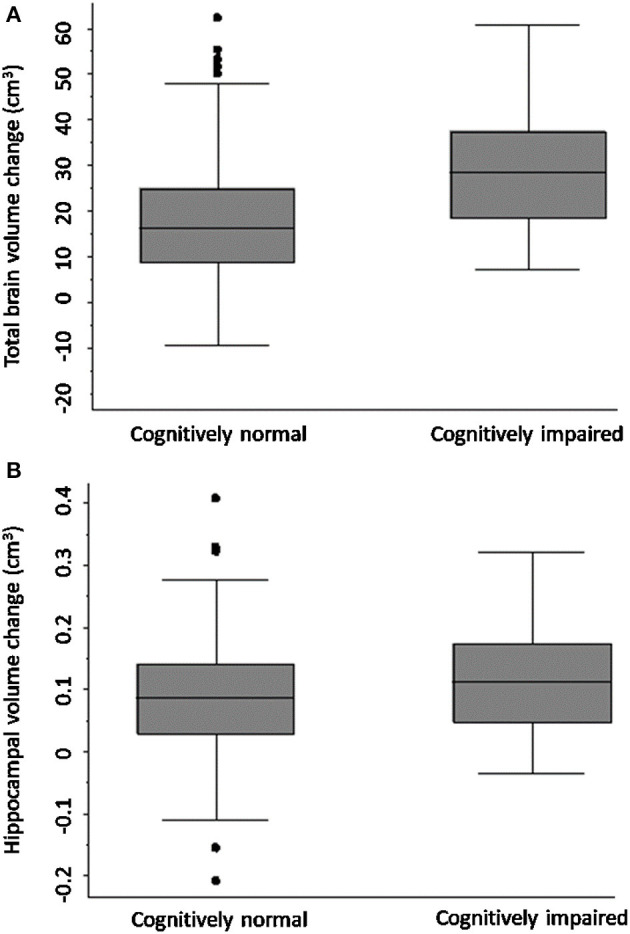
Stroke survivors are at high risk of dementia, associated with increasing age and vascular burden and with pre-existing cognitive impairment, older age. Brain atrophy patterns are recognised as signatures of neurodegenerative conditions, but the natural history of brain atrophy after stroke remains poorly described. We sought to determine whether stroke survivors who were cognitively normal at time of stroke had greater total brain (TBV) and hippocampal volume (HV) loss over 3 years than controls. We examined whether stroke survivors who were cognitively impaired (CI) at 3 months following their stroke had greater brain volume loss than cognitively normal (CN) stroke participants over the next 3 years. Cognition And Neocortical Volume After Stroke (CANVAS) study is a multi-centre cohort study of first-ever or recurrent adult ischaemic stroke participants compared to age- and sex-matched community controls. Participants were followed with MRI and cognitive assessments over 3 years and were free of a history of cognitive impairment or decline at inclusion. Our primary outcome measure was TBV change between 3 months and 3 years; secondary outcomes were TBV and HV change comparing CI and CN participants. We investigated associations between group status and brain volume change using a baseline-volume adjusted linear regression model with robust standard error. Ninety-three stroke (26 women, 66.7 ± 12 years) and 39 control participants (15 women, 68.7 ± 7 years) were available at 3 years. TBV loss in stroke patients was greater than controls: stroke mean () = 20.3 cm ± SD 14.8 cm; controls = 14.2 cm ± SD 13.2 cm; [adjusted mean difference 7.88 95%CI (2.84, 12.91) -value = 0.002]. TBV decline was greater in those stroke participants who were cognitively impaired ( = 30.7 cm; SD = 14.2 cm) at 3 months ( = 19.6 cm; SD = 13.8 cm); [adjusted mean difference 10.42; 95%CI (3.04, 17.80), -value = 0.006]. No statistically significant differences in HV change were observed. Ischaemic stroke survivors exhibit greater neurodegeneration compared to stroke-free controls. Brain atrophy is greater in stroke participants who were cognitively impaired early after their stroke. Early cognitive impairment was associated greater subsequent atrophy, reflecting the combined impacts of stroke and vascular brain burden. Atrophy rates could serve as a useful biomarker for trials testing interventions to reduce post-stroke secondary neurodegeneration. http://www.clinicaltrials.gov, identifier: NCT02205424.
中风幸存者患痴呆症的风险很高,这与年龄增长、血管负担加重以及先前存在的认知障碍、高龄有关。脑萎缩模式被认为是神经退行性疾病的特征,但中风后脑萎缩的自然史仍描述甚少。我们试图确定中风时认知正常的中风幸存者在3年内的全脑(TBV)和海马体体积(HV)损失是否比对照组更大。我们研究了中风后3个月时认知受损(CI)的中风幸存者在接下来3年中脑体积损失是否比认知正常(CN)的中风参与者更大。中风后认知与新皮质体积(CANVAS)研究是一项多中心队列研究,将首次发生或复发性成人缺血性中风参与者与年龄和性别匹配的社区对照组进行比较。参与者在3年内接受了MRI和认知评估,入组时无认知障碍或衰退史。我们的主要结局指标是3个月至3年期间的TBV变化;次要结局是比较CI和CN参与者的TBV和HV变化。我们使用具有稳健标准误的基线体积调整线性回归模型研究了组状态与脑体积变化之间的关联。3年后有93名中风患者(26名女性,66.7±12岁)和39名对照参与者(15名女性,68.7±7岁)可供研究。中风患者的TBV损失大于对照组:中风组平均值()=20.3 cm±标准差14.8 cm;对照组=14.2 cm±标准差13.2 cm;[调整后平均差异7.88 95%置信区间(2.84,12.91)-值=0.002]。3个月时认知受损的中风参与者(=30.7 cm;标准差=14.2 cm)的TBV下降更大(=19.6 cm;标准差=13.8 cm);[调整后平均差异10.42;95%置信区间(3.04,17.80),-值=0.006]。未观察到HV变化有统计学显著差异。与无中风的对照组相比,缺血性中风幸存者表现出更大的神经退行性变。中风后早期认知受损的中风参与者脑萎缩更严重。早期认知障碍与随后更大的萎缩相关,反映了中风和脑血管负担的综合影响。萎缩率可作为测试减少中风后继发性神经退行性变干预措施的试验的有用生物标志物。http://www.clinicaltrials.gov,标识符:NCT02205424。