• 文献检索
  • 文档翻译
  • 深度研究
  • 学术资讯
  • Suppr Zotero 插件Zotero 插件
  • 邀请有礼
  • 套餐&价格
  • 历史记录
应用&插件
Suppr Zotero 插件Zotero 插件浏览器插件Mac 客户端Windows 客户端微信小程序
定价
高级版会员购买积分包购买API积分包
服务
文献检索文档翻译深度研究API 文档MCP 服务
关于我们
关于 Suppr公司介绍联系我们用户协议隐私条款
关注我们

Suppr 超能文献

核心技术专利:CN118964589B侵权必究
粤ICP备2023148730 号-1Suppr @ 2026

文献检索

告别复杂PubMed语法,用中文像聊天一样搜索,搜遍4000万医学文献。AI智能推荐,让科研检索更轻松。

立即免费搜索

文件翻译

保留排版,准确专业,支持PDF/Word/PPT等文件格式,支持 12+语言互译。

免费翻译文档

深度研究

AI帮你快速写综述,25分钟生成高质量综述,智能提取关键信息,辅助科研写作。

立即免费体验

急性 COVID-19 感染患者的心室变化:世界超声心动图学会联盟(WASE-COVID)研究的随访。

Ventricular Changes in Patients with Acute COVID-19 Infection: Follow-up of the World Alliance Societies of Echocardiography (WASE-COVID) Study.

机构信息

University of Chicago, Chicago, Illinois.

Ultromics, Oxford, United Kingdom.

出版信息

J Am Soc Echocardiogr. 2022 Mar;35(3):295-304. doi: 10.1016/j.echo.2021.10.015. Epub 2021 Nov 6.

DOI:10.1016/j.echo.2021.10.015
PMID:34752928
原文链接:https://pmc.ncbi.nlm.nih.gov/articles/PMC8572036/
Abstract

BACKGROUND

COVID-19 infection is known to cause a wide array of clinical chronic sequelae, but little is known regarding the long-term cardiac complications. We aim to report echocardiographic follow-up findings and describe the changes in left (LV) and right ventricular (RV) function that occur following acute infection.

METHODS

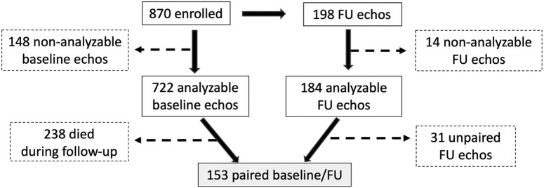
Patients enrolled in the World Alliance Societies of Echocardiography-COVID study with acute COVID-19 infection were asked to return for a follow-up transthoracic echocardiogram. Overall, 198 returned at a mean of 129 days of follow-up, of which 153 had paired baseline and follow-up images that were analyzable, including LV volumes, ejection fraction (LVEF), and longitudinal strain (LVLS). Right-sided echocardiographic parameters included RV global longitudinal strain, RV free wall strain, and RV basal diameter. Paired echocardiographic parameters at baseline and follow-up were compared for the entire cohort and for subgroups based on the baseline LV and RV function.

RESULTS

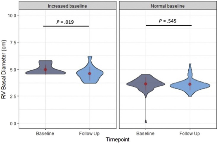
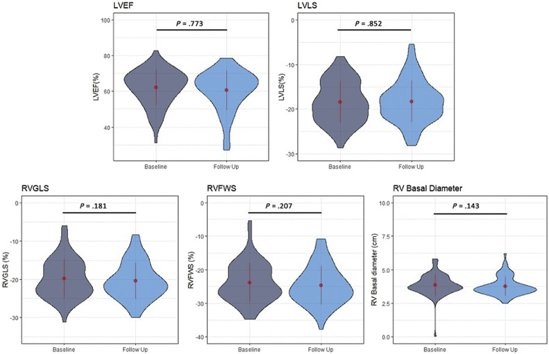
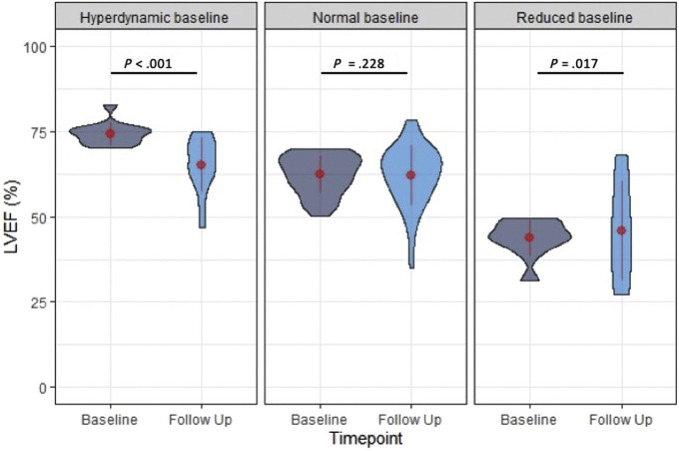
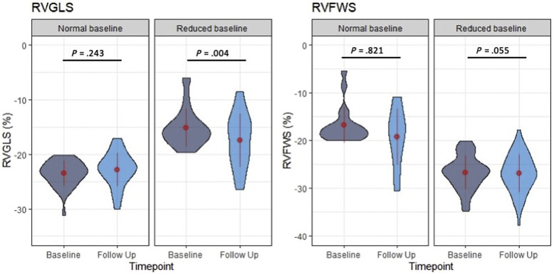
For the entire cohort, echocardiographic markers of LV and RV function at follow-up were not significantly different from baseline (all P > .05). Patients with hyperdynamic LVEF at baseline (>70%), had a significant reduction of LVEF at follow-up (74.3% ± 3.1% vs 64.4% ± 8.1%, P < .001), while patients with reduced LVEF at baseline (<50%) had a significant increase (42.5% ± 5.9% vs 49.3% ± 13.4%, P = .02), and those with normal LVEF had no change. Patients with normal LVLS (<-18%) at baseline had a significant reduction of LVLS at follow-up (-21.6% ± 2.6% vs -20.3% ± 4.0%, P = .006), while patients with impaired LVLS at baseline had a significant improvement at follow-up (-14.5% ± 2.9% vs -16.7% ± 5.2%, P < .001). Patients with abnormal RV global longitudinal strain (>-20%) at baseline had significant improvement at follow-up (-15.2% ± 3.4% vs -17.4% ± 4.9%, P = .004). Patients with abnormal RV basal diameter (>4.5 cm) at baseline had significant improvement at follow-up (4.9 ± 0.7 cm vs 4.6 ± 0.6 cm, P = .019).

CONCLUSIONS

Overall, there were no significant changes over time in the LV and RV function of patients recovering from COVID-19 infection. However, differences were observed according to baseline LV and RV function, which may reflect recovery from the acute myocardial injury occurring in the acutely ill. Left ventricular and RV function tends to improve in those with impaired baseline function, while it tends to decrease in those with hyperdynamic LV or normal RV function.

摘要

背景

已知 COVID-19 感染可引起广泛的临床慢性后遗症,但对长期心脏并发症知之甚少。我们旨在报告超声心动图随访结果,并描述急性感染后左(LV)和右心室(RV)功能的变化。

方法

在世界超声心动图学会联盟 COVID 研究中招募了患有急性 COVID-19 感染的患者,并要求他们返回进行随访经胸超声心动图检查。共有 198 名患者在平均 129 天的随访中返回,其中 153 名患者具有基线和随访的配对图像,可进行分析,包括 LV 容积、射血分数(LVEF)和纵向应变(LVLS)。右侧超声心动图参数包括 RV 整体纵向应变、RV 游离壁应变和 RV 基底直径。比较整个队列和基于基线 LV 和 RV 功能的亚组的基线和随访时的配对超声心动图参数。

结果

对于整个队列,LV 和 RV 功能的超声心动图标志物在随访时与基线相比没有显著差异(均 P >.05)。基线时射血分数高(>70%)的患者,随访时射血分数明显降低(74.3%±3.1% vs 64.4%±8.1%,P<.001),而基线时射血分数降低(<50%)的患者射血分数显著增加(42.5%±5.9% vs 49.3%±13.4%,P=.02),而射血分数正常的患者没有变化。基线时 LVLS 正常(<-18%)的患者,随访时 LVLS 明显下降(-21.6%±2.6% vs -20.3%±4.0%,P=.006),而基线时 LVLS 受损的患者随访时明显改善(-14.5%±2.9% vs -16.7%±5.2%,P<.001)。基线时 RV 整体纵向应变异常(>-20%)的患者,随访时明显改善(-15.2%±3.4% vs -17.4%±4.9%,P=.004)。基线时 RV 基底直径异常(>4.5 cm)的患者,随访时明显改善(4.9±0.7 cm vs 4.6±0.6 cm,P=.019)。

结论

总体而言,从 COVID-19 感染中恢复的患者的 LV 和 RV 功能在随访期间没有明显变化。然而,根据基线 LV 和 RV 功能观察到差异,这可能反映了急性疾病中发生的急性心肌损伤的恢复。基线功能受损的患者的左心室和 RV 功能趋于改善,而 LV 射血分数高或 RV 功能正常的患者的左心室和 RV 功能趋于下降。

https://cdn.ncbi.nlm.nih.gov/pmc/blobs/2452/8572036/9deb4b38436c/gr6_lrg.jpg
https://cdn.ncbi.nlm.nih.gov/pmc/blobs/2452/8572036/533519bcdc67/gr1_lrg.jpg
https://cdn.ncbi.nlm.nih.gov/pmc/blobs/2452/8572036/41c083ae0bcb/gr2_lrg.jpg
https://cdn.ncbi.nlm.nih.gov/pmc/blobs/2452/8572036/165ab5d7491c/gr3_lrg.jpg
https://cdn.ncbi.nlm.nih.gov/pmc/blobs/2452/8572036/d820b074ba45/gr4_lrg.jpg
https://cdn.ncbi.nlm.nih.gov/pmc/blobs/2452/8572036/d9622610d8c6/gr5_lrg.jpg
https://cdn.ncbi.nlm.nih.gov/pmc/blobs/2452/8572036/9deb4b38436c/gr6_lrg.jpg

相似文献

1
Ventricular Changes in Patients with Acute COVID-19 Infection: Follow-up of the World Alliance Societies of Echocardiography (WASE-COVID) Study.急性 COVID-19 感染患者的心室变化:世界超声心动图学会联盟(WASE-COVID)研究的随访。
J Am Soc Echocardiogr. 2022 Mar;35(3):295-304. doi: 10.1016/j.echo.2021.10.015. Epub 2021 Nov 6.
2
Echocardiographic Correlates of In-Hospital Death in Patients with Acute COVID-19 Infection: The World Alliance Societies of Echocardiography (WASE-COVID) Study.急性 COVID-19 感染患者住院期间死亡的超声心动图相关性:世界超声心动图联盟协会(WASE-COVID)研究。
J Am Soc Echocardiogr. 2021 Aug;34(8):819-830. doi: 10.1016/j.echo.2021.05.010. Epub 2021 May 21.
3
Left and right ventricular dysfunction in patients with COVID-19-associated myocardial injury.COVID-19 相关心肌损伤患者的左右心室功能障碍。
Infection. 2021 Jun;49(3):491-500. doi: 10.1007/s15010-020-01572-8. Epub 2021 Jan 30.
4
Right ventricular global longitudinal strain provides prognostic value incremental to left ventricular ejection fraction in patients with heart failure.右心室整体纵向应变提供的预后价值优于心力衰竭患者的左心室射血分数。
J Am Soc Echocardiogr. 2014 Jul;27(7):726-32. doi: 10.1016/j.echo.2014.02.007. Epub 2014 Mar 26.
5
Similarities and Differences in Left Ventricular Size and Function among Races and Nationalities: Results of the World Alliance Societies of Echocardiography Normal Values Study.不同种族和民族之间左心室大小和功能的异同:世界超声心动图联盟协会正常数值研究的结果。
J Am Soc Echocardiogr. 2019 Nov;32(11):1396-1406.e2. doi: 10.1016/j.echo.2019.08.012.
6
Human versus Artificial Intelligence-Based Echocardiographic Analysis as a Predictor of Outcomes: An Analysis from the World Alliance Societies of Echocardiography COVID Study.基于人工智能的超声心动图分析与人预测结局的比较:来自世界超声心动图联盟 COVID 研究的分析。
J Am Soc Echocardiogr. 2022 Dec;35(12):1226-1237.e7. doi: 10.1016/j.echo.2022.07.004. Epub 2022 Jul 19.
7
Evolution of right and left ventricle routine and speckle-tracking echocardiography in patients recovering from coronavirus disease 2019: a longitudinal study.新冠肺炎患者右、左心室常规及斑点追踪超声心动图演变:一项纵向研究。
Eur Heart J Cardiovasc Imaging. 2022 Jul 21;23(8):1055-1065. doi: 10.1093/ehjci/jeab190.
8
Echocardiographic Characteristics of Left and Right Ventricular Longitudinal Function in Patients With a History of Cardiac Surgery.有心脏手术史患者左、右心室纵向功能的超声心动图特征
Am J Cardiol. 2024 Jan 15;211:72-78. doi: 10.1016/j.amjcard.2023.10.045. Epub 2023 Oct 22.
9
Risk Stratification of Future Left Ventricular Dysfunction for Patients with Indications for Right Ventricular Pacing due to Bradycardia.因心动过缓而有右心室起搏指征患者未来左心室功能障碍的风险分层
Int Heart J. 2017 Oct 21;58(5):724-730. doi: 10.1536/ihj.16-415. Epub 2017 Sep 30.
10
Cardiologic Manifestations in Omicron-Type Versus Wild-Type COVID-19: A Systematic Echocardiographic Study.奥密克戎变异株与野生型新冠病毒所致心血管系统表现的系统超声心动图研究。
J Am Heart Assoc. 2023 Feb 7;12(3):e027188. doi: 10.1161/JAHA.122.027188. Epub 2023 Jan 25.

引用本文的文献

1
Cardiac structure and function 1.5 years after COVID-19: results from the EPILOC study.新冠病毒感染后1.5年的心脏结构和功能:EPILOC研究结果
Infection. 2025 Feb 24. doi: 10.1007/s15010-025-02481-4.
2
A deeper look at long-term effects of COVID-19 on myocardial function in survivors with no prior heart diseases: a GRADE approach systematic review and meta-analysis.深入研究新冠病毒病对无既往心脏病幸存者心肌功能的长期影响:采用GRADE方法进行的系统评价和荟萃分析
Front Cardiovasc Med. 2024 Nov 19;11:1458389. doi: 10.3389/fcvm.2024.1458389. eCollection 2024.
3
Cardiac Function and Structure before and after Mild SARS-CoV-2 Infection in Elite Athletes Using Biventricular and Left Atrial Strain.
使用双心室和左心房应变评估精英运动员轻度感染新型冠状病毒2后心脏功能和结构的变化
Biomedicines. 2024 Oct 11;12(10):2310. doi: 10.3390/biomedicines12102310.
4
Serial evaluation of biventricular function in COVID-19 recovered patients using speckle tracking echocardiography.应用斑点追踪超声心动图对 COVID-19 康复患者的双心室功能进行连续评估。
Indian Heart J. 2024 Jul-Aug;76(4):297-302. doi: 10.1016/j.ihj.2024.08.002. Epub 2024 Aug 26.
5
Clinical utility of ambulatory ECG monitoring and 2D-ventricular strain for evaluation of post-COVID-19 ventricular arrhythmia.动态心电图监测和二维心室应变在评估新冠病毒后室性心律失常中的临床应用。
BMC Cardiovasc Disord. 2024 Aug 16;24(1):429. doi: 10.1186/s12872-024-03982-0.
6
Association Between Baseline Echocardiographic Parameters and Acute Coronavirus Disease 2019 Infection in Hospitalized Patients.住院患者基线超声心动图参数与2019年冠状病毒病急性感染之间的关联
Cureus. 2024 Mar 3;16(3):e55432. doi: 10.7759/cureus.55432. eCollection 2024 Mar.
7
Comprehensive temporal analysis of right ventricular function and pulmonary haemodynamics in mechanically ventilated COVID-19 ARDS patients.机械通气的新型冠状病毒肺炎急性呼吸窘迫综合征患者右心室功能和肺血流动力学的综合时间分析
Ann Intensive Care. 2024 Feb 12;14(1):25. doi: 10.1186/s13613-024-01241-1.
8
New-Onset Atrial Fibrillation in the Critically Ill COVID-19 Patients Hospitalized in the Intensive Care Unit.入住重症监护病房的危重症COVID-19患者新发房颤
J Clin Med. 2023 Nov 8;12(22):6989. doi: 10.3390/jcm12226989.
9
Assessment of myocardial function and cardiac performance using left ventricular global longitudinal strain in athletes after COVID-19: a follow-up study.新冠疫情后运动员左心室整体纵向应变评估心肌功能和心脏性能:一项随访研究
Front Cardiovasc Med. 2023 Oct 9;10:1240278. doi: 10.3389/fcvm.2023.1240278. eCollection 2023.
10
Serial echocardiographic evaluation of COVID-19 patients without prior history of structural heart disease: a 1-year follow-up CRACoV-HHS study.对无结构性心脏病既往史的COVID-19患者进行系列超声心动图评估:一项为期1年的CRACoV-HHS随访研究。
Front Cardiovasc Med. 2023 Sep 13;10:1230669. doi: 10.3389/fcvm.2023.1230669. eCollection 2023.