• 文献检索
  • 文档翻译
  • 深度研究
  • 学术资讯
  • Suppr Zotero 插件Zotero 插件
  • 邀请有礼
  • 套餐&价格
  • 历史记录
应用&插件
Suppr Zotero 插件Zotero 插件浏览器插件Mac 客户端Windows 客户端微信小程序
定价
高级版会员购买积分包购买API积分包
服务
文献检索文档翻译深度研究API 文档MCP 服务
关于我们
关于 Suppr公司介绍联系我们用户协议隐私条款
关注我们

Suppr 超能文献

核心技术专利:CN118964589B侵权必究
粤ICP备2023148730 号-1Suppr @ 2026

文献检索

告别复杂PubMed语法,用中文像聊天一样搜索,搜遍4000万医学文献。AI智能推荐,让科研检索更轻松。

立即免费搜索

文件翻译

保留排版,准确专业,支持PDF/Word/PPT等文件格式,支持 12+语言互译。

免费翻译文档

深度研究

AI帮你快速写综述,25分钟生成高质量综述,智能提取关键信息,辅助科研写作。

立即免费体验

膝关节负重及早期弯曲状态下基于 MRI 的半月板 3D 形态变化。

The MRI-based 3D morphologic changes of knee meniscus under knee weight-bearing and early flexion conditions.

机构信息

Department of Orthopedics, China-Japan Union Hospital of Jilin University, Changchun, Jilin Province, People's Republic of China.

Department of Orthopedics, The Second Hospital of Jilin University, Changchun, Jilin Province, People's Republic of China.

出版信息

Sci Rep. 2021 Nov 11;11(1):22122. doi: 10.1038/s41598-021-01531-9.

DOI:10.1038/s41598-021-01531-9
PMID:34764374
原文链接:https://pmc.ncbi.nlm.nih.gov/articles/PMC8586250/
Abstract

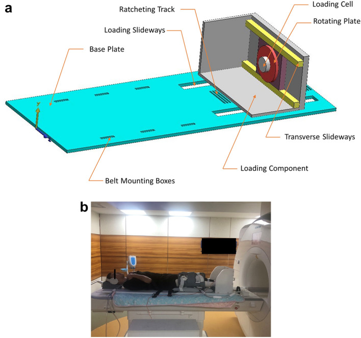
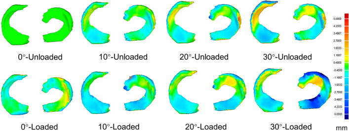
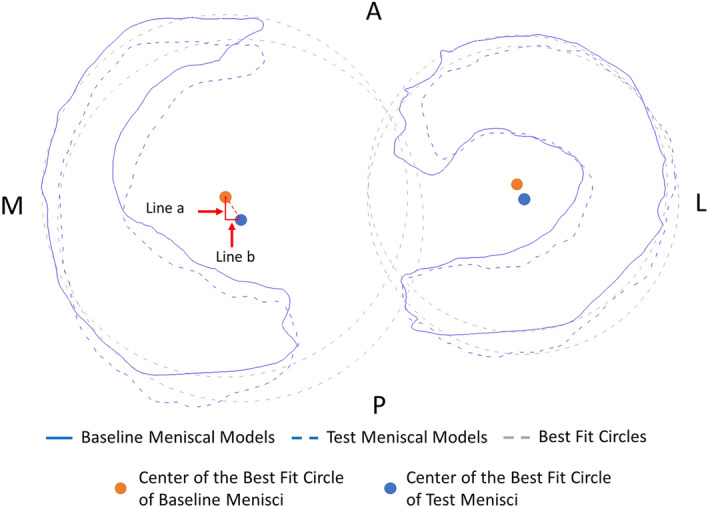
There are few studies investigate morphologic changes of knee meniscus in vivo mechanical loading and three-dimensions (3D) deformation and displacement of the whole meniscus between in vivo mechanical loading and unloading conditions are still unclear. To investigate the displacements and 3D morphological changes of the menisci under knee weight-bearing and early flexion conditions in healthy adults using a Magnetic Resonance Imaging (MRI)-compatible loading device (a 3.0 T MR imaging system) combined with a newly developed 3D comparison technique. Fifteen healthy volunteers were recruited in this cross-sectional observational study. Each subject underwent MRIs of their dominant right knee in eight different scanning conditions using a 3.0-T MRI scanner with a custom-made MRI-compatible loading device. The knee meniscus images were 3D reconstructed, and dimensional comparisons were made for each meniscal model with baseline (0°-unloaded model). The morphologic changes of the meniscal-anterior horn (AH), body (BD), and posterior horn (PH) regions were expressed as mean positive and negative deviations. The displacements were further investigated, and the meniscal extrusions of different subregions were measured. The morphologic changing patterns of human meniscus under loading and flexions were presented using 3D chromatic maps. The bilateral menisci were generally shifting laterally and posteriorly in most flexion angles and were changing medially and anteriorly under fully extended knee loading conditions. The mean deviations were more significant with loading at 0° of knee flexion, while the PH region in the lateral side changed further posteriorly with loading in 30° flexion. Most of the differences were not significant in other flexion angles between loading conditions. The extrusion of meniscus's medial body was greater in full extension compared to any other flexing angles. Mechanical loading can significantly deform the menisci in knee extension; however, this effect is limited during knee flexion. Current study can be used as a reference for the evaluations of the integrity in meniscal functions.

摘要

目前,很少有研究调查体内机械负荷下膝关节半月板的形态变化,以及在体内负荷和卸载条件下整个半月板的三维(3D)变形和位移。本研究旨在使用磁共振成像(MRI)兼容的加载设备(3.0T MRI 成像系统)结合新开发的 3D 比较技术,研究健康成年人膝关节负重和早期弯曲条件下半月板的位移和 3D 形态变化。本研究共纳入 15 名健康志愿者。每位受试者在 3.0T MRI 扫描仪上使用定制的 MRI 兼容加载设备,对其优势右膝进行 8 种不同扫描条件的 MRI 检查。对半月板图像进行三维重建,并与基线(0°-未加载模型)比较每个半月板模型的尺寸。以正负偏差的平均值表示半月板前角(AH)、体部(BD)和后角(PH)区域的形态变化。进一步研究了半月板的位移,并测量了不同亚区的半月板突出。使用 3D 彩色图谱呈现了加载和弯曲下人体半月板的形态变化模式。在大多数弯曲角度下,双侧半月板普遍向外侧和后侧移位,在完全伸展的膝关节加载条件下向内侧和前侧移位。在膝关节弯曲 0°时,半月板的平均偏差更为显著,而在 30°弯曲时,外侧的 PH 区进一步向后移位。在其他弯曲角度下,加载条件之间的差异大多不显著。与任何其他弯曲角度相比,半月板体部的内侧突出更大。膝关节伸展时,机械负荷可显著改变半月板的形态;然而,在膝关节弯曲时,这种影响是有限的。本研究可为评估半月板功能的完整性提供参考。

https://cdn.ncbi.nlm.nih.gov/pmc/blobs/b6fb/8586250/cd6588ef22d6/41598_2021_1531_Fig3_HTML.jpg
https://cdn.ncbi.nlm.nih.gov/pmc/blobs/b6fb/8586250/48f60ccb35fc/41598_2021_1531_Fig1_HTML.jpg
https://cdn.ncbi.nlm.nih.gov/pmc/blobs/b6fb/8586250/b8674a1f0e05/41598_2021_1531_Fig2_HTML.jpg
https://cdn.ncbi.nlm.nih.gov/pmc/blobs/b6fb/8586250/cd6588ef22d6/41598_2021_1531_Fig3_HTML.jpg
https://cdn.ncbi.nlm.nih.gov/pmc/blobs/b6fb/8586250/48f60ccb35fc/41598_2021_1531_Fig1_HTML.jpg
https://cdn.ncbi.nlm.nih.gov/pmc/blobs/b6fb/8586250/b8674a1f0e05/41598_2021_1531_Fig2_HTML.jpg
https://cdn.ncbi.nlm.nih.gov/pmc/blobs/b6fb/8586250/cd6588ef22d6/41598_2021_1531_Fig3_HTML.jpg

相似文献

1
The MRI-based 3D morphologic changes of knee meniscus under knee weight-bearing and early flexion conditions.膝关节负重及早期弯曲状态下基于 MRI 的半月板 3D 形态变化。
Sci Rep. 2021 Nov 11;11(1):22122. doi: 10.1038/s41598-021-01531-9.
2
Kinematic change of the meniscus and the tibiofemoral joint space in asymptomatic volunteers using a wide bore 3T closed MRI system.使用大孔径3T封闭式MRI系统对无症状志愿者半月板及胫股关节间隙的运动学变化进行研究。
Skeletal Radiol. 2015 Oct;44(10):1441-51. doi: 10.1007/s00256-015-2187-4. Epub 2015 Jun 16.
3
In vivo assessment of weight-bearing knee flexion reveals compartment-specific alterations in meniscal slope.体内评估承重膝关节屈曲可揭示半月板斜率的特定间室改变。
Arthroscopy. 2013 Oct;29(10):1653-60. doi: 10.1016/j.arthro.2013.07.260. Epub 2013 Aug 30.
4
Utilization of Transtibial Centralization Suture Best Minimizes Extrusion and Restores Tibiofemoral Contact Mechanics for Anatomic Medial Meniscal Root Repairs in a Cadaveric Model.在尸体模型中,利用经胫骨中央化缝合术可最大程度减少挤出并恢复胫股接触力学,从而实现解剖内侧半月板根部修复。
Am J Sports Med. 2019 Jun;47(7):1591-1600. doi: 10.1177/0363546519844250. Epub 2019 May 15.
5
Predicting meniscal tear stability across knee-joint flexion using finite-element analysis.基于有限元分析预测膝关节弯曲时半月板撕裂的稳定性。
Knee Surg Sports Traumatol Arthrosc. 2019 Jan;27(1):206-214. doi: 10.1007/s00167-018-5090-4. Epub 2018 Aug 10.
6
MRI-based kinematics of the menisci through full knee range of motion.基于磁共振成像的半月板在全膝关节活动范围内的运动学研究。
J Orthop Surg (Hong Kong). 2021 May-Aug;29(2):23094990211017349. doi: 10.1177/23094990211017349.
7
Medial meniscus extrusion increases with age and BMI and is depending on different loading conditions.内侧半月板挤出随着年龄和 BMI 的增加而增加,并取决于不同的加载条件。
Knee Surg Sports Traumatol Arthrosc. 2018 Aug;26(8):2282-2288. doi: 10.1007/s00167-018-4885-7. Epub 2018 Mar 6.
8
Investigation of extrusion of the medial meniscus under full weight-loading conditions using upright weight-loading magnetic resonance imaging and ultrasonography.使用直立负重磁共振成像和超声检查对全负重条件下内侧半月板挤出情况的研究。
J Orthop Sci. 2020 Jul;25(4):652-657. doi: 10.1016/j.jos.2019.09.009. Epub 2019 Oct 5.
9
Normal and transplanted lateral knee menisci: evaluation of extrusion using magnetic resonance imaging and ultrasound.正常及移植后的外侧膝关节半月板:利用磁共振成像和超声评估半月板挤出情况
Knee Surg Sports Traumatol Arthrosc. 2004 Sep;12(5):411-9. doi: 10.1007/s00167-004-0500-1. Epub 2004 May 14.
10
Displacement of the medial meniscus within the passive motion characteristics of the human knee joint: an RSA study in human cadaver knees.人体膝关节被动运动特征下内侧半月板的移位:人体尸体膝关节的RSA研究
Knee Surg Sports Traumatol Arthrosc. 2005 May;13(4):287-92. doi: 10.1007/s00167-004-0511-y. Epub 2004 Aug 11.

引用本文的文献

1
Biomechanics of horizontal meniscus tear and healing during knee flexion: Finite element analysis.膝关节屈曲时水平半月板撕裂与愈合的生物力学:有限元分析
Mechanobiol Med. 2025 Mar 13;3(2):100128. doi: 10.1016/j.mbm.2025.100128. eCollection 2025 Jun.
2
Significantly smaller lateral extrusion was observed within 24 weeks after all-inside suture repairs of radial tear in the middle segment of lateral meniscus compared to inside-out repairs.与由外向内修复相比,在外侧半月板中段放射状撕裂的全内缝合修复术后24周内观察到明显更小的外侧挤压。
J Exp Orthop. 2024 Oct 15;11(4):e70041. doi: 10.1002/jeo2.70041. eCollection 2024 Oct.
3

本文引用的文献

1
Morphological Analysis of Normal Meniscus on Magnetic Resonance Imaging (MRI)-Based Three-Dimensional Reconstruction Models in Healthy Chinese Adults.基于磁共振成像(MRI)三维重建模型的健康中国成年人正常半月板形态学分析
Med Sci Monit. 2020 Oct 15;26:e927101. doi: 10.12659/MSM.927101.
2
Magnetic resonance imaging of human knee joint functionality under variable compressive in-situ loading and axis alignment.可变原位压缩载荷和轴线对齐条件下人体膝关节功能的磁共振成像
J Mech Behav Biomed Mater. 2020 Oct;110:103890. doi: 10.1016/j.jmbbm.2020.103890. Epub 2020 May 30.
3
Comparison of two different on-shelf femoral stems for Crowe type IV developmental dysplasia of the hip.
Qualitative Magnetic Resonance Imaging Assessment of the Semimembranosus Tendon in Patients with Medial Meniscal Tears.
内侧半月板撕裂患者半膜肌腱的定性磁共振成像评估
Diagnostics (Basel). 2024 Sep 5;14(17):1962. doi: 10.3390/diagnostics14171962.
4
The relation between meniscal dynamics and tibiofemoral kinematics.半月板动力学与胫股关节运动学之间的关系。
Sci Rep. 2024 Apr 17;14(1):8829. doi: 10.1038/s41598-024-59265-3.
5
Extrusion and meniscal mobility evaluation in case of ramp lesion injury: a biomechanical feasibility study by 7T magnetic resonance imaging and digital volume correlation.斜坡损伤情况下的半月板挤出和活动度评估:一项通过7T磁共振成像和数字体积相关技术的生物力学可行性研究
Front Bioeng Biotechnol. 2024 Jan 5;11:1289290. doi: 10.3389/fbioe.2023.1289290. eCollection 2023.
6
Toe-out gait inhibits medial meniscus extrusion associated with the second peak of knee adduction moment during gait in patients with knee osteoarthritis.在膝骨关节炎患者中,外翻步态可抑制与步态期间膝关节内收力矩第二个峰值相关的内侧半月板挤压。
Asia Pac J Sports Med Arthrosc Rehabil Technol. 2023 Aug 24;33:13-19. doi: 10.1016/j.asmart.2023.08.001. eCollection 2023 Jul.
7
Evaluation of meniscus extrusion during stair ambulation in healthy volunteers using dynamic ultrasonography: a feasibility study.使用动态超声评估健康志愿者在楼梯活动时半月板挤出:一项可行性研究。
J Med Ultrason (2001). 2023 Oct;50(4):541-549. doi: 10.1007/s10396-023-01348-1. Epub 2023 Aug 11.
8
Unique patterns of medial meniscus extrusion during walking and its association with limb kinematics in patients with knee osteoarthritis.膝关节骨关节炎患者行走时内侧半月板挤出的独特模式及其与肢体运动学的关系。
Sci Rep. 2023 Aug 2;13(1):12513. doi: 10.1038/s41598-023-39715-0.
9
Decrease in medial meniscal extrusion after physical therapy to improve knee pain and range of motion in patients with knee osteoarthritis: A retrospective study.物理治疗后内侧半月板突出减少可改善膝骨关节炎患者的膝关节疼痛和活动范围:一项回顾性研究。
PLoS One. 2022 Nov 30;17(11):e0277628. doi: 10.1371/journal.pone.0277628. eCollection 2022.
两种不同的用于Crowe IV型髋关节发育不良的股骨柄假体的比较。
J Int Med Res. 2020 Aug;48(8):300060520947888. doi: 10.1177/0300060520947888.
4
An MRI-compatible varus-valgus loading device for whole-knee joint functionality assessment based on compartmental compression: a proof-of-concept study.一种基于腔室压缩的全膝关节功能评估的 MRI 兼容内外翻加载装置:概念验证研究。
MAGMA. 2020 Dec;33(6):839-854. doi: 10.1007/s10334-020-00844-6. Epub 2020 Apr 20.
5
Magnetic resonance imaging (MRI) studies of knee joint under mechanical loading: Review.膝关节在机械负荷下的磁共振成像(MRI)研究:综述。
Magn Reson Imaging. 2020 Jan;65:27-36. doi: 10.1016/j.mri.2019.09.007. Epub 2019 Oct 25.
6
Investigation of extrusion of the medial meniscus under full weight-loading conditions using upright weight-loading magnetic resonance imaging and ultrasonography.使用直立负重磁共振成像和超声检查对全负重条件下内侧半月板挤出情况的研究。
J Orthop Sci. 2020 Jul;25(4):652-657. doi: 10.1016/j.jos.2019.09.009. Epub 2019 Oct 5.
7
No dynamic extrusion of the medial meniscus in ultrasound examination in patients with confirmed root tear lesion.在确诊根撕裂病变的患者的超声检查中,未见内侧半月板的动态外突。
Knee Surg Sports Traumatol Arthrosc. 2019 Oct;27(10):3311-3317. doi: 10.1007/s00167-018-5341-4. Epub 2019 Jan 3.
8
Medial meniscus extrusion increases with age and BMI and is depending on different loading conditions.内侧半月板挤出随着年龄和 BMI 的增加而增加,并取决于不同的加载条件。
Knee Surg Sports Traumatol Arthrosc. 2018 Aug;26(8):2282-2288. doi: 10.1007/s00167-018-4885-7. Epub 2018 Mar 6.
9
Clinical platform for understanding the relationship between joint contact mechanics and articular cartilage changes after meniscal surgery.用于理解半月板手术后关节接触力学与关节软骨变化之间关系的临床平台。
J Orthop Res. 2017 Mar;35(3):600-611. doi: 10.1002/jor.23365. Epub 2017 Feb 13.
10
Loaded versus unloaded magnetic resonance imaging (MRI) of the knee: Effect on meniscus extrusion in healthy volunteers and patients with osteoarthritis.膝关节负重与非负重磁共振成像(MRI):对健康志愿者和骨关节炎患者半月板挤出的影响
Eur J Radiol Open. 2016 May 20;3:100-7. doi: 10.1016/j.ejro.2016.05.002. eCollection 2016.