Department of Biomechanics, Graduate School of Biomedical and Health Sciences, Hiroshima University, 1-2-3 Kasumi, Minami-ku, Hiroshima, 734-8553, Japan.
Department of Orthopaedic Surgery, Faculty of Medicine, Kagawa University, Kagawa, Japan.
Sci Rep. 2023 Aug 2;13(1):12513. doi: 10.1038/s41598-023-39715-0.
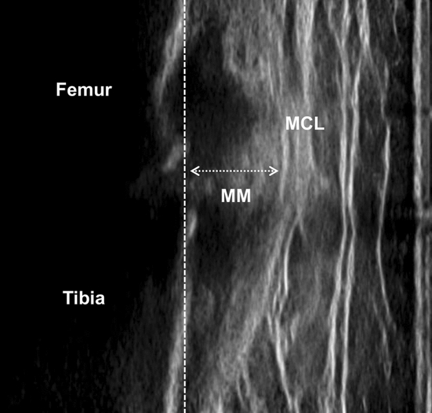
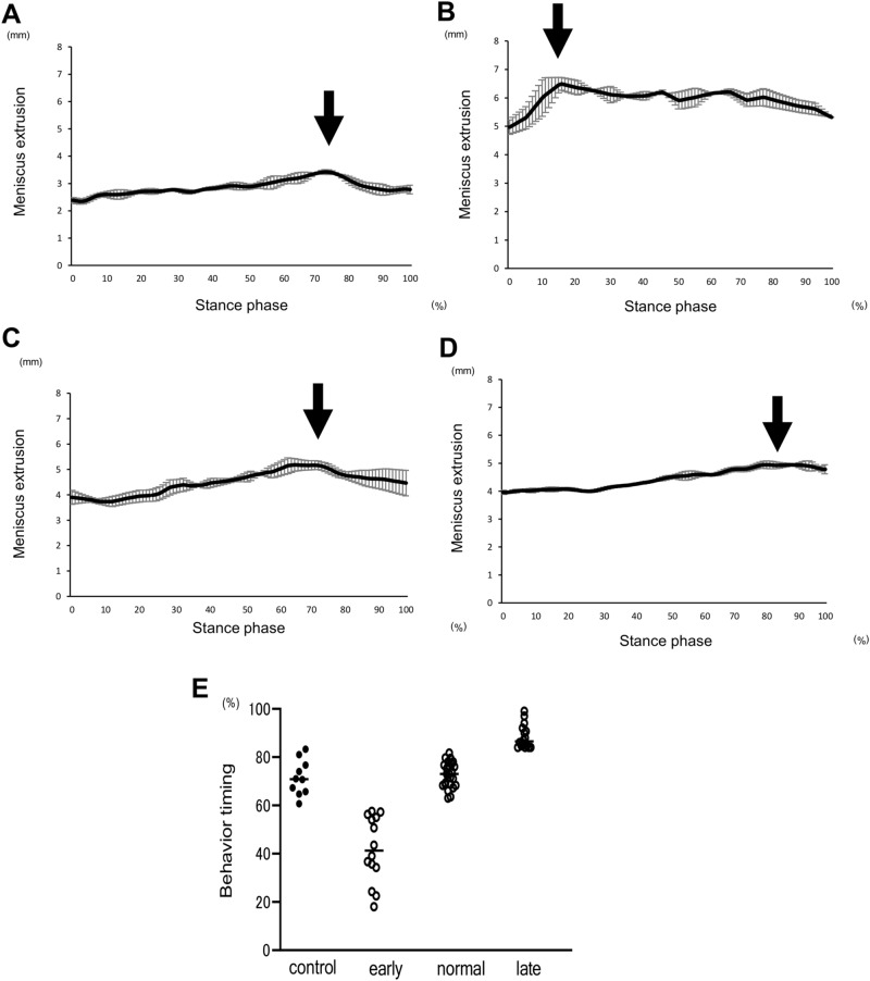
Medial meniscus extrusion (MME) is exacerbated by repeated mechanical stress. Various factors would affect MME; however, there is limited information about the behaviour of the medial meniscus during walking in patients with knee osteoarthritis (KOA). This study aimed to investigate the pattern of MME during walking and its association with limb biomechanics in patients with KOA. Fifty-five patients with KOA and ten older adult volunteers as a control group were involved in this study. The MME and limb biomechanics during walking were evaluated simultaneously by ultrasound and a motion analysis system, respectively. The waveform was constructed from the values of MME, and the point showing the highest value of MME was identified during the gait cycle. According to the peak timing of MME in the waveform, the pattern of the waveform was evaluated and compared to the control group. Lateral thrust, knee adduction moment (KAM), and flexion moment were obtained from motion analysis, and their association with the MME was evaluated. The patients with KOA demonstrated unique peak timing during walking. Compared to the control group, there were three groups of MME waveforms, early (< 59%), normal (60-83%), and late (> 84%) from the peak timing in the gait cycle. The pattern of MME waveform in early, normal, and late groups was correlated with the first KAM and lateral thrust, second KAM, and knee flexion moment, respectively. A unique MME pattern during walking was demonstrated, and these patterns were associated with limb biomechanics in patients with KOA.
内侧半月板挤压(MME)可因反复机械应力而加剧。许多因素都会影响 MME;然而,关于膝骨关节炎(KOA)患者在行走过程中内侧半月板的行为,信息有限。本研究旨在探讨 KOA 患者在行走过程中 MME 的模式及其与肢体生物力学的关系。本研究共纳入 55 例 KOA 患者和 10 名老年志愿者作为对照组。通过超声和运动分析系统分别评估行走过程中的 MME 和肢体生物力学。根据 MME 值构建波形,并确定步态周期中 MME 值最高的点。根据波形中 MME 的峰值时间,评估并比较其与对照组的波形模式。从运动分析中获得横向推力、膝内收力矩(KAM)和弯曲力矩,并评估其与 MME 的关系。KOA 患者在行走过程中表现出独特的峰值时间。与对照组相比,从步态周期中的峰值时间来看,MME 有三组波形,早期(<59%)、正常(60-83%)和晚期(>84%)。早期、正常和晚期 MME 波形的模式分别与第一 KAM 和横向推力、第二 KAM 和膝关节弯曲力矩相关。在行走过程中表现出独特的 MME 模式,这些模式与 KOA 患者的肢体生物力学有关。