Whi Wonseok, Choi Hongyoon, Paeng Jin Chul, Cheon Gi Jeong, Kang Keon Wook, Lee Dong Soo
Department of Molecular Medicine and Biopharmaceutical Sciences, Seoul National University, Seoul, 03080, Republic of Korea.
Department of Nuclear Medicine, Seoul National University College of Medicine, 101 Daehak-ro, Jongno-gu, Seoul, 03080, Republic of Korea.
EJNMMI Phys. 2021 Nov 14;8(1):79. doi: 10.1186/s40658-021-00424-0.
The whole brain is often covered in [F]Fluorodeoxyglucose positron emission tomography ([F]FDG-PET) in oncology patients, but the covered brain abnormality is typically screened by visual interpretation without quantitative analysis in clinical practice. In this study, we aimed to develop a fully automated quantitative interpretation pipeline of brain volume from an oncology PET image.
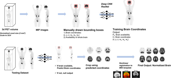
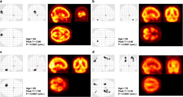
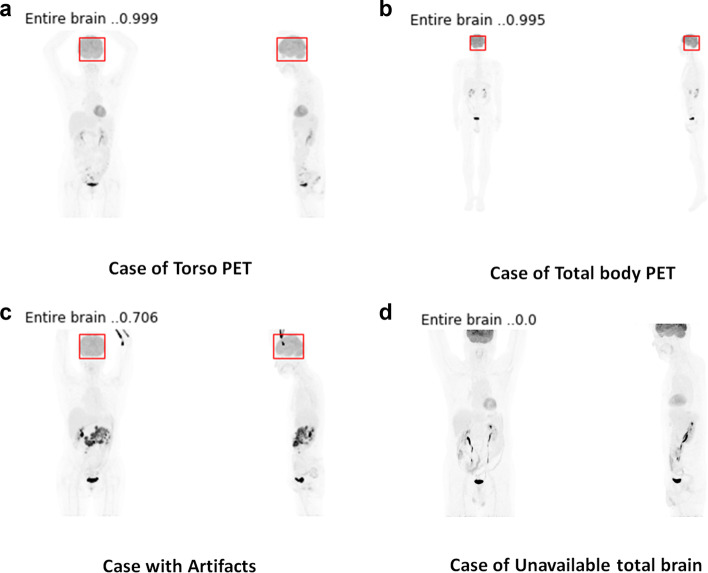
We retrospectively collected 500 oncologic [F]FDG-PET scans for training and validation of the automated brain extractor. We trained the model for extracting brain volume with two manually drawn bounding boxes on maximal intensity projection images. ResNet-50, a 2-D convolutional neural network (CNN), was used for the model training. The brain volume was automatically extracted using the CNN model and spatially normalized. For validation of the trained model and an application of this automated analytic method, we enrolled 24 subjects with small cell lung cancer (SCLC) and performed voxel-wise two-sample T test for automatic detection of metastatic lesions.
The deep learning-based brain extractor successfully identified the existence of whole-brain volume, with an accuracy of 98% for the validation set. The performance of extracting the brain measured by the intersection-over-union of 3-D bounding boxes was 72.9 ± 12.5% for the validation set. As an example of the application to automatically identify brain abnormality, this approach successfully identified the metastatic lesions in three of the four cases of SCLC patients with brain metastasis.
Based on the deep learning-based model, extraction of the brain volume from whole-body PET was successfully performed. We suggest this fully automated approach could be used for the quantitative analysis of brain metabolic patterns to identify abnormalities during clinical interpretation of oncologic PET studies.
在肿瘤患者中,[F]氟脱氧葡萄糖正电子发射断层扫描([F]FDG-PET)常覆盖全脑,但在临床实践中,所覆盖的脑异常通常通过视觉解读进行筛查,而不进行定量分析。在本研究中,我们旨在开发一种从肿瘤PET图像中全自动定量解读脑容量的流程。
我们回顾性收集了500例肿瘤[F]FDG-PET扫描图像,用于训练和验证自动脑提取器。我们在最大强度投影图像上使用两个手动绘制的边界框训练用于提取脑容量的模型。使用二维卷积神经网络(CNN)ResNet-50进行模型训练。使用CNN模型自动提取脑容量并进行空间归一化。为了验证训练好的模型并应用这种自动分析方法,我们招募了24例小细胞肺癌(SCLC)患者,并进行体素级双样本T检验以自动检测转移灶。
基于深度学习的脑提取器成功识别出全脑容量的存在,验证集的准确率为98%。通过三维边界框的交并比测量的脑提取性能在验证集中为72.9±12.5%。作为自动识别脑异常应用的一个例子,该方法成功识别出4例脑转移SCLC患者中的3例的转移灶。
基于深度学习模型,成功从全身PET中提取了脑容量。我们建议这种全自动方法可用于脑代谢模式的定量分析,以在肿瘤PET研究的临床解读过程中识别异常。