Laliotis Nikolaos, Chrysanthou Chrysanthos, Konstandinidis Panagiotis, Papadopoulou Elisavet
Department of Orthopaedics, Inter Balkan Medical Center, Thessaloniki, Greece.
Department of Radiology, Inter Balkan Medical Center, Thessaloniki, Greece.
J Orthop Case Rep. 2021 Jul;11(7):90-93. doi: 10.13107/jocr.2021.v11.i07.2332.
Solitary osteochondromas are extremely rare in the bones of the foot. In the growing skeleton, few cases affecting the metatarsals and the talus have been reported. At present, there have been no reports of osteochondromas affecting the cuneiforms.
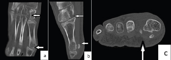
We report the case of a 13-year-old male patient. He presented with marked prominences in the plantar surface of his left foot and pain while participating in sporting activities. Radiological examination with X-rays, computed tomography (CT) scan, and magnetic resonance imaging revealed two solitary osteochondromas growing from the medial cuneiform and the head of the 1st metatarsal. The patient was treated surgically by excision of the osteochondromas. Histological examination confirmed the diagnosis of osteochondromas. He had an uneventful recovery and returned to his sporting activities.
Solitary osteochondroma can present in the cuneiform and metatarsal of a growing adolescent. CT scan is useful for the accurate diagnosis and surgical removal of the tumor.
孤立性骨软骨瘤在足部骨骼中极为罕见。在生长中的骨骼中,仅有少数累及跖骨和距骨的病例报道。目前,尚无关于骨软骨瘤累及楔骨的报道。
我们报告一例13岁男性患者。他因左脚足底明显隆起且在参加体育活动时疼痛而就诊。X线、计算机断层扫描(CT)及磁共振成像等影像学检查显示,有两个孤立性骨软骨瘤分别从内侧楔骨和第一跖骨头长出。该患者接受了骨软骨瘤切除术。组织学检查确诊为骨软骨瘤。他恢复顺利,重返体育活动。
孤立性骨软骨瘤可出现在生长中的青少年的楔骨和跖骨。CT扫描有助于准确诊断及手术切除肿瘤。