Zhang Jie, Wang Haixia, Yao Linlin, Zhao Peng, Wu Xiaoyan
Department of Nutrition, Shandong Provincial Hospital, Cheeloo College of Medicine, Shandong University, Jinan, China.
Department of Nutrition, Shandong Provincial Hospital Affiliated to Shandong First Medical University, Jinan, China.
Ann Transl Med. 2021 Oct;9(20):1520. doi: 10.21037/atm-21-5005.
Previous studies have confirmed that MicroRNA (miRNA) is a key regulator exhibiting different effects in human liver fibrosis. However, the function of miR-34a in liver fibrosis has not been reported. Hence, this study aimed to investigate the regulatory mechanism of miR-34a in liver fibrosis.
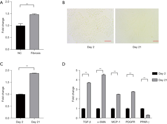
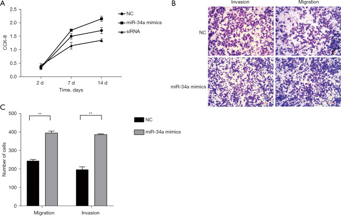
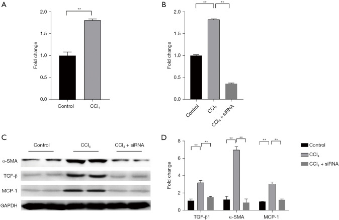
The expression of miR-34a was measured in fibrosis tissues via the quantitative real-time PCR (qRT-PCR) assay. Subsequently, 30 male C57BL/6J mice were divided into control and treatment groups and used to establish animal models of liver fibrosis to explore the expression level of miR-34a. Moreover, Cell Counting Kit 8 (CCK-8) and transwell assays were preformed to identify the regulatory mechanism of miR-34a in cells. The effect of miR-34a on the activity of transforming growth factor-β (TGF-β) pathway was observed by western blot.
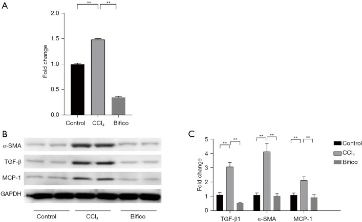
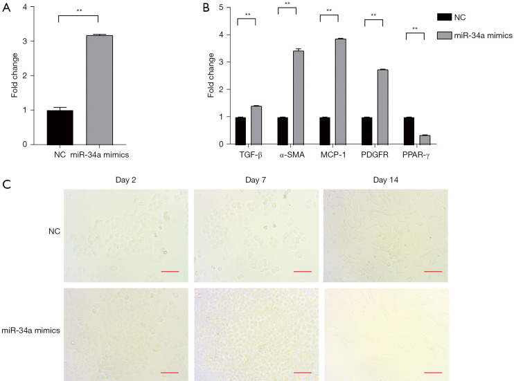
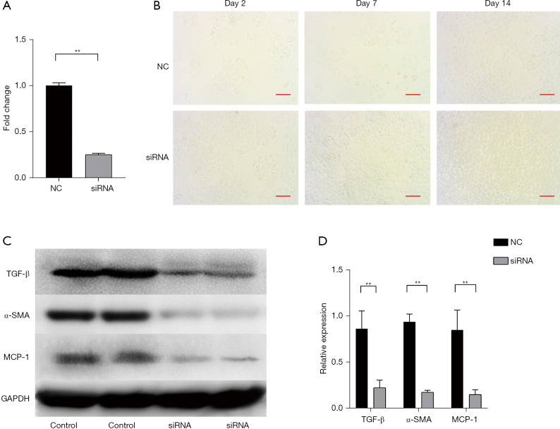
Up-regulation of miR-34a was detected in fibrosis cells. Moreover, the cellular phenotype was suppressed by miR-34a down-regulation in a primary culture of hepatic stellate cells (HSCs). Besides, it was found that increased miR-34a could significantly promote the invasion and migration of HSCs. Moreover, miR-34a activates HSCs through transforming TGF-β, α-smooth muscle actin (α-SMA), and Monocyte chemoattractant protein-1 (MCP-1), which further affects liver fibrosis.
MiR-34a promotes the fibrosis of HSCs as well as cell proliferation, migration, and invasion.
先前的研究已证实,微小RNA(miRNA)是一种关键调节因子,在人类肝纤维化中发挥着不同作用。然而,miR-34a在肝纤维化中的功能尚未见报道。因此,本研究旨在探讨miR-34a在肝纤维化中的调控机制。
通过定量实时聚合酶链反应(qRT-PCR)检测纤维化组织中miR-34a的表达。随后,将30只雄性C57BL/6J小鼠分为对照组和治疗组,用于建立肝纤维化动物模型,以探究miR-34a的表达水平。此外,进行细胞计数试剂盒8(CCK-8)和transwell实验,以确定miR-34a在细胞中的调控机制。通过蛋白质免疫印迹法观察miR-34a对转化生长因子-β(TGF-β)信号通路活性的影响。
在纤维化细胞中检测到miR-34a上调。此外,在肝星状细胞(HSCs)原代培养中,miR-34a下调可抑制细胞表型。此外,发现miR-34a增加可显著促进HSCs的侵袭和迁移。此外,miR-34a通过转化TGF-β、α-平滑肌肌动蛋白(α-SMA)和单核细胞趋化蛋白-1(MCP-1)激活HSCs,进而影响肝纤维化。
MiR-34a促进HSCs纤维化以及细胞增殖、迁移和侵袭。