Department of Diagnostic and Interventional radiology, All India Institute of Medical Sciences, Jodhpur, Rajasthan, India.
Department of Diagnostic and Interventional radiology, All India Institute of Medical Sciences, Jodhpur, Rajasthan, India.
Curr Probl Diagn Radiol. 2022 Jan-Feb;51(1):112-120. doi: 10.1067/j.cpradiol.2021.09.004. Epub 2021 Nov 3.
Rhino-orbital-cerebral mucormycosis has emerged as a major opportunistic infection in patients with COVID-19. High clinical suspicion and prompt imaging are crucial for early diagnosis and management. Our study evaluates imaging characteristics of patients with COVID-19 associated Rhino-orbital-cerebral Mucormycosis (CA-ROCM) in a tertiary care hospital in India.
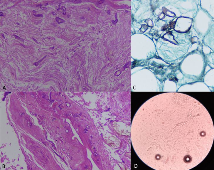
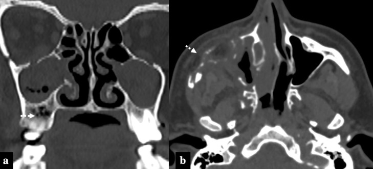
A retrospective analysis of clinical and imaging data of patients with CA-ROCM who presented between December 2020 to June 2021 was performed. All patients had microbiologically or histologically proven sino-nasal mucormycosis along with documented SARS-CoV-2 positive RT-PCR test and/or classical lung imaging features of COVID-19 infection. The extent of sinus involvement, bony erosions, extra-sinus soft tissue extension, orbital-intracranial invasion, perineural spread, and vascular complications were assessed.
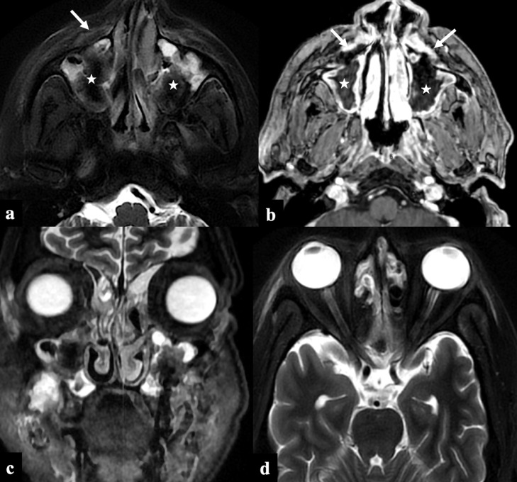
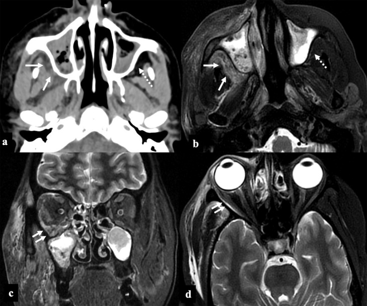
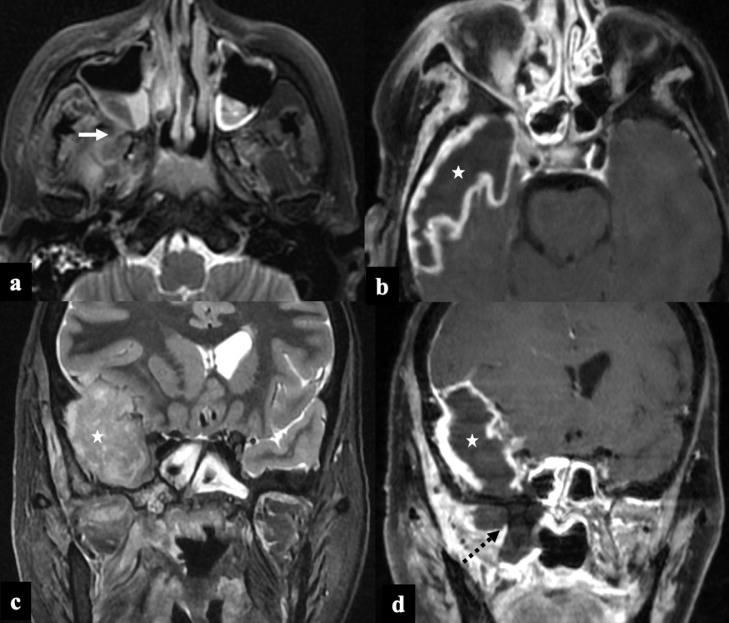
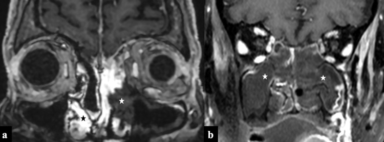
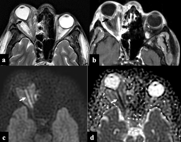
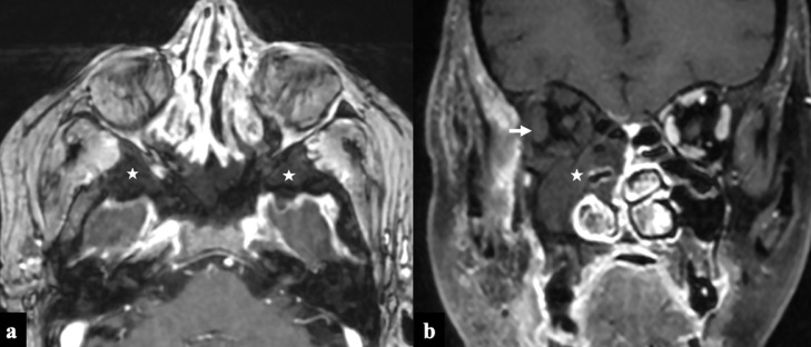
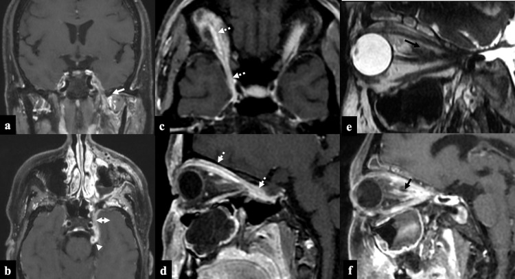
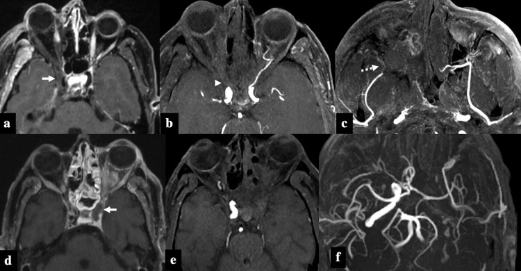
Fifty patients were included for the final analysis. Diabetes was the most common associated comorbidity. Seven patients presented with stage I disease, 18 patients with stage II, and 25 patients with stage III disease. The stage of disease showed a positive statistical correlation with HbA1c levels using Pearson's correlation. The common imaging features were "Black turbinate sign" and nonenhancing sino-nasal mucosa (82%), orbital involvement (76%), and diffusion restriction in the optic nerve (24%). Intracranial involvement was seen as perineural extension into the brain (42%), cerebritis (30%), and internal carotid artery involvement (16%).
CA-ROCM is an acute invasive fungal sinusitis with an aggressive clinical course. Black-turbinate sign and peri-antral soft tissue infiltration are early features, whereas extra-nasal tissue infarction, optic nerve diffusion restriction, and vascular invasion are seen with advanced disease.
COVID-19 患者中,鼻-眶-脑毛霉菌病已成为一种主要的机会性感染。高临床怀疑和及时的影像学检查对于早期诊断和治疗至关重要。我们的研究评估了印度一家三级保健医院 COVID-19 相关鼻-眶-脑毛霉菌病(CA-ROCM)患者的影像学特征。
对 2020 年 12 月至 2021 年 6 月期间出现 CA-ROCM 的患者的临床和影像学数据进行回顾性分析。所有患者均经微生物学或组织学证实存在鼻窦毛霉菌病,同时有记录的 SARS-CoV-2 阳性 RT-PCR 检测和/或 COVID-19 感染的典型肺部影像学特征。评估了鼻窦受累程度、骨侵蚀、鼻窦外软组织延伸、眼眶-颅内侵犯、神经周围扩散和血管并发症。
最终有 50 例患者纳入分析。糖尿病是最常见的合并症。7 例患者为 I 期疾病,18 例为 II 期疾病,25 例为 III 期疾病。疾病分期与 Pearson 相关性分析显示 HbA1c 水平呈正相关。常见的影像学特征包括“黑鼻甲征”和无增强的鼻窦黏膜(82%)、眼眶受累(76%)和视神经弥散受限(24%)。颅内受累表现为神经周围向脑内延伸(42%)、脑实质炎(30%)和颈内动脉受累(16%)。
CA-ROCM 是一种侵袭性急性真菌性鼻窦炎,具有侵袭性的临床病程。黑鼻甲征和前鼻窦软组织浸润是早期特征,而鼻窦外组织梗死、视神经弥散受限和血管侵犯则见于晚期疾病。