Sanchez-Varo Raquel, Sanchez-Mejias Elisabeth, Fernandez-Valenzuela Juan Jose, De Castro Vanessa, Mejias-Ortega Marina, Gomez-Arboledas Angela, Jimenez Sebastian, Sanchez-Mico Maria Virtudes, Trujillo-Estrada Laura, Moreno-Gonzalez Ines, Baglietto-Vargas David, Vizuete Marisa, Davila Jose Carlos, Vitorica Javier, Gutierrez Antonia
Departamento Biologia Celular, Genetica y Fisiologia, Instituto de Investigacion Biomedica de Malaga-IBIMA, Facultad de Ciencias, Universidad de Málaga, Málaga, Spain.
Centro de Investigación Biomedica en Red Sobre Enfermedades Neurodegenerativas (CIBERNED), Madrid, Spain.
Front Neurosci. 2021 Nov 4;15:752594. doi: 10.3389/fnins.2021.752594. eCollection 2021.
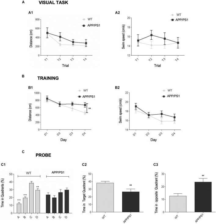
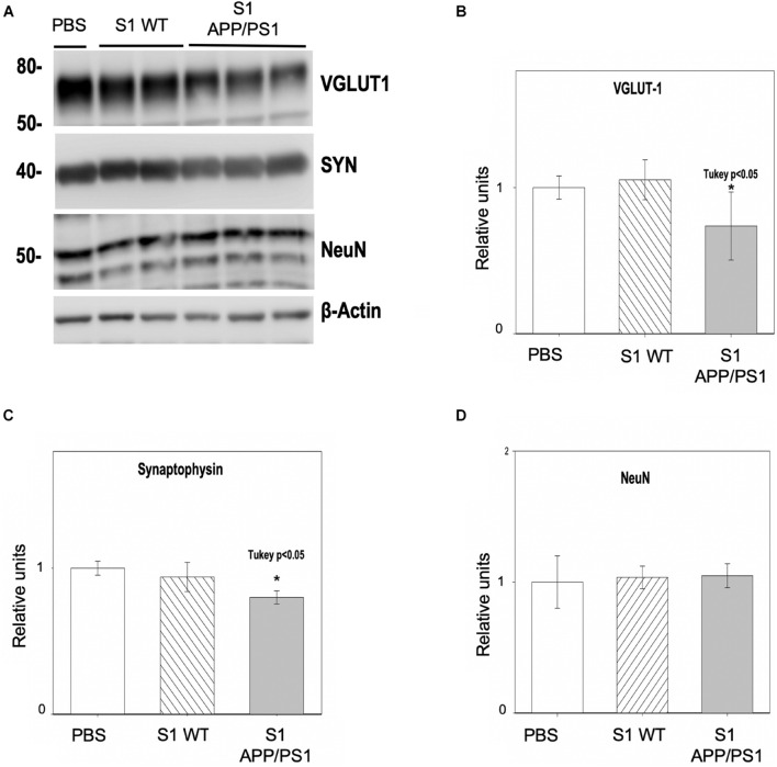
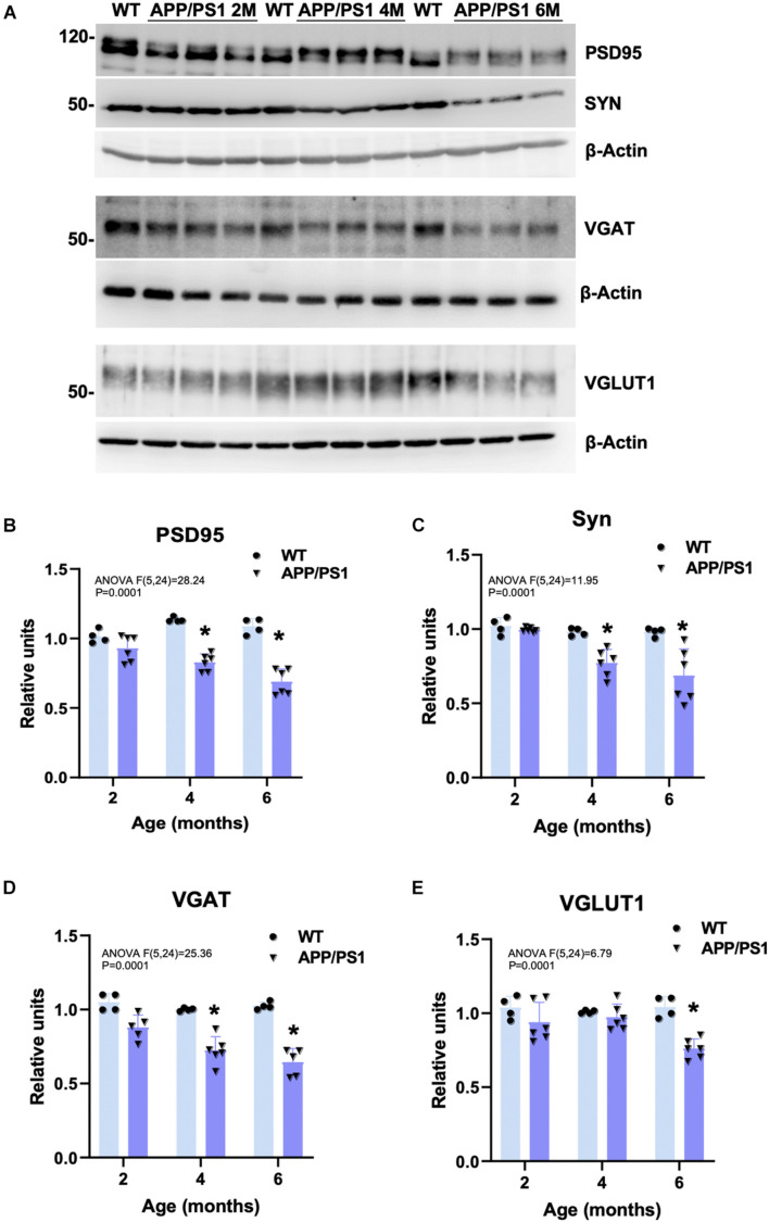
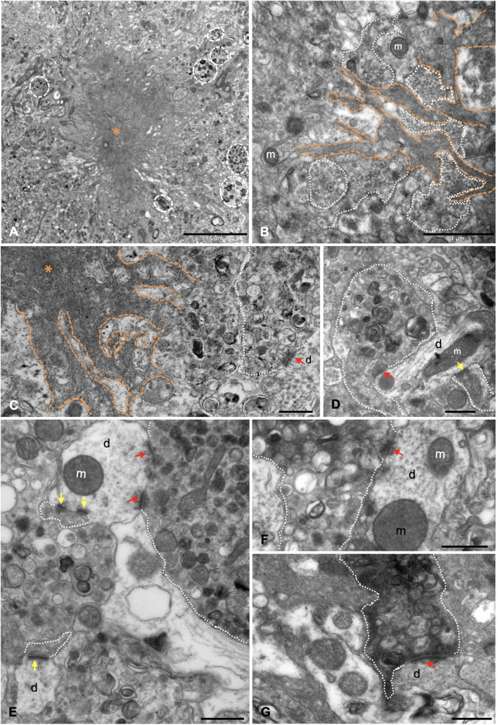
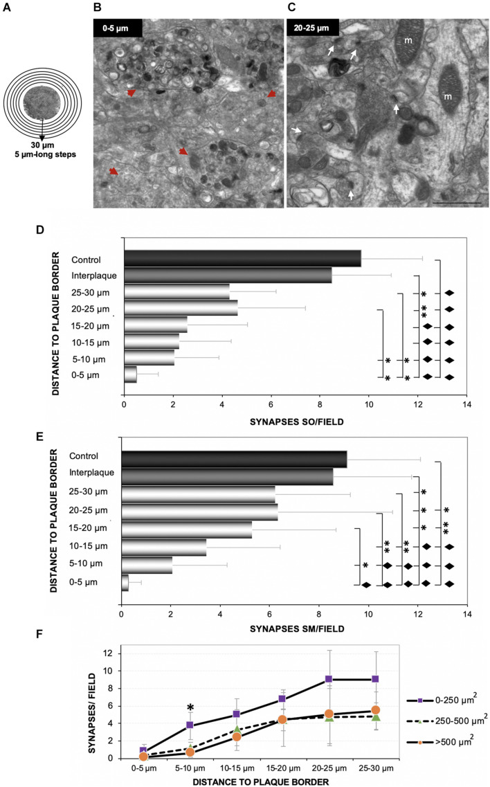
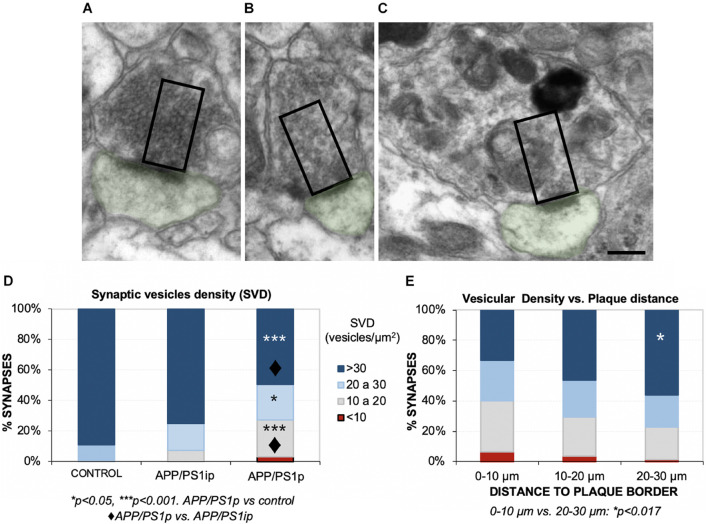
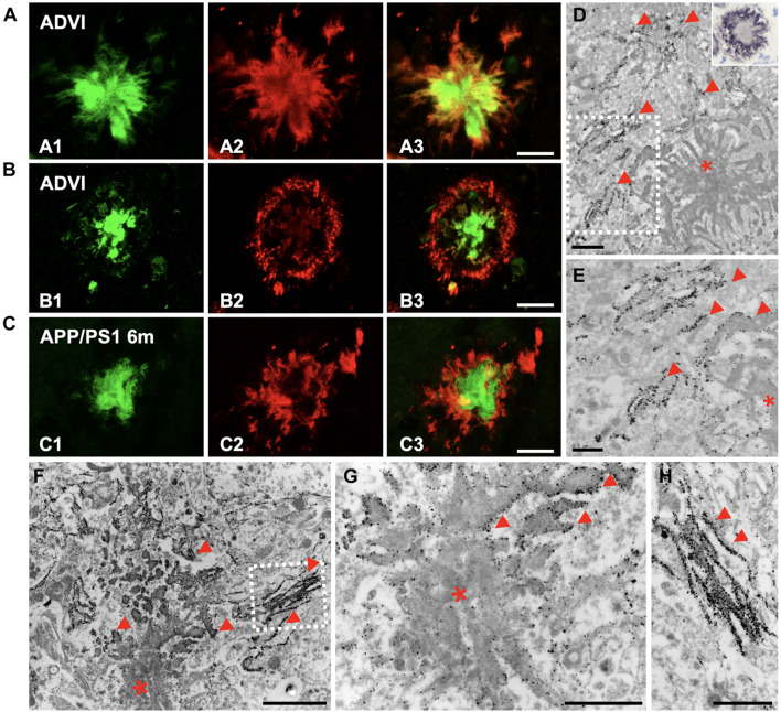
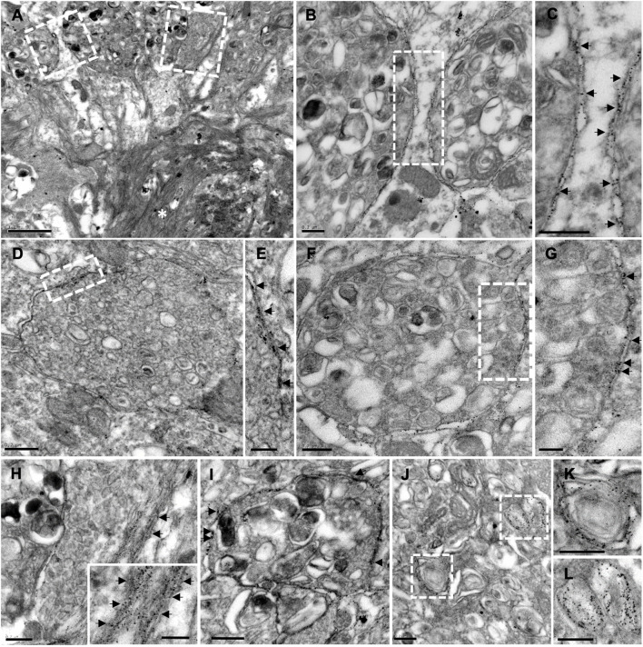
Alzheimer's disease (AD) is a devastating neurodegenerative disorder characterized by initial memory impairments that progress to dementia. In this sense, synaptic dysfunction and loss have been established as the pathological features that best correlate with the typical early cognitive decline in this disease. At the histopathological level, AD brains typically exhibit intraneuronal neurofibrillary tangles (NFTs) along with the accumulation of amyloid-beta (Abeta) peptides in the form of extracellular deposits. Specifically, the oligomeric soluble forms of Abeta are considered the most synaptotoxic species. In addition, neuritic plaques are Abeta deposits surrounded by activated microglia and astroglia cells together with abnormal swellings of neuronal processes named dystrophic neurites. These periplaque aberrant neurites are mostly presynaptic elements and represent the first pathological indicator of synaptic dysfunction. In terms of losing synaptic proteins, the hippocampus is one of the brain regions most affected in AD patients. In this work, we report an early decline in spatial memory, along with hippocampal synaptic changes, in an amyloidogenic APP/PS1 transgenic model. Quantitative electron microscopy revealed a spatial synaptotoxic pattern around neuritic plaques with significant loss of periplaque synaptic terminals, showing rising synapse loss close to the border, especially in larger plaques. Moreover, dystrophic presynapses were filled with autophagic vesicles in detriment of the presynaptic vesicular density, probably interfering with synaptic function at very early synaptopathological disease stages. Electron immunogold labeling showed that the periphery of amyloid plaques, and the associated dystrophic neurites, was enriched in Abeta oligomers supporting an extracellular location of the synaptotoxins. Finally, the incubation of primary neurons with soluble fractions derived from 6-month-old APP/PS1 hippocampus induced significant loss of synaptic proteins, but not neuronal death. Indeed, this preclinical transgenic model could serve to investigate therapies targeted at initial stages of synaptic dysfunction relevant to the prodromal and early AD.
阿尔茨海默病(AD)是一种毁灭性的神经退行性疾病,其特征是最初的记忆障碍会发展为痴呆。从这个意义上说,突触功能障碍和丧失已被确认为与该疾病典型早期认知衰退最相关的病理特征。在组织病理学水平上,AD 大脑通常表现为神经元内神经原纤维缠结(NFTs),以及以细胞外沉积物形式存在的β淀粉样蛋白(Aβ)肽的积累。具体而言,Aβ 的寡聚可溶性形式被认为是最具突触毒性的种类。此外,神经炎性斑块是被激活的小胶质细胞和星形胶质细胞包围的 Aβ 沉积物,以及被称为营养不良性神经突的神经元突起的异常肿胀。这些斑块周围的异常神经突大多是突触前元件,代表突触功能障碍的第一个病理指标。就突触蛋白丧失而言,海马体是 AD 患者中受影响最严重的脑区之一。在这项研究中,我们报告了在淀粉样蛋白生成的 APP/PS1 转基因模型中,空间记忆的早期衰退以及海马体突触变化。定量电子显微镜显示神经炎性斑块周围存在空间突触毒性模式,斑块周围突触终末显著丧失,显示靠近边界处突触丧失增加,尤其是在较大的斑块中。此外,营养不良性突触前膜充满自噬囊泡,损害了突触前囊泡密度,可能在突触病理疾病的非常早期阶段干扰突触功能。电子免疫金标记显示淀粉样斑块的周边以及相关的营养不良性神经突富含 Aβ 寡聚体,支持突触毒素的细胞外定位。最后,用来自 6 个月大的 APP/PS1 海马体可溶性组分孵育原代神经元会导致突触蛋白显著丧失,但不会导致神经元死亡。事实上,这个临床前转基因模型可用于研究针对与前驱期和早期 AD 相关的突触功能障碍初始阶段的疗法。