Mejía-Chávez Sara, Venebra-Muñoz Arturo, García-García Fabio, Corona-Morales Aleph Alejandro, Orozco-Vargas Arturo Enrique
Laboratorio de Neurobiología de la Adicción y Plasticidad Cerebral, Facultad de Ciencias, Universidad Autónoma del Estado de Mexico, Toluca, Mexico.
Laboratorio de Biología de Sueño, Instituto de Ciencias de la Salud, Universidad Veracruzana, Xalapa, Mexico.
Front Behav Neurosci. 2021 Nov 4;15:651263. doi: 10.3389/fnbeh.2021.651263. eCollection 2021.
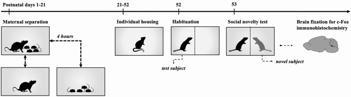
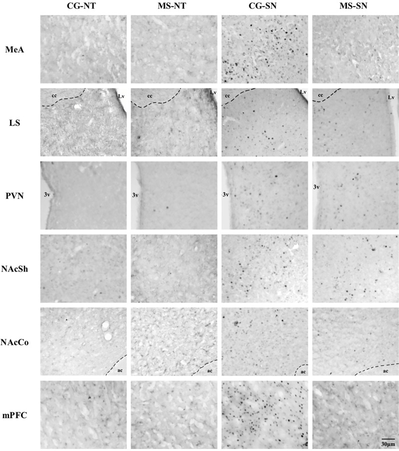
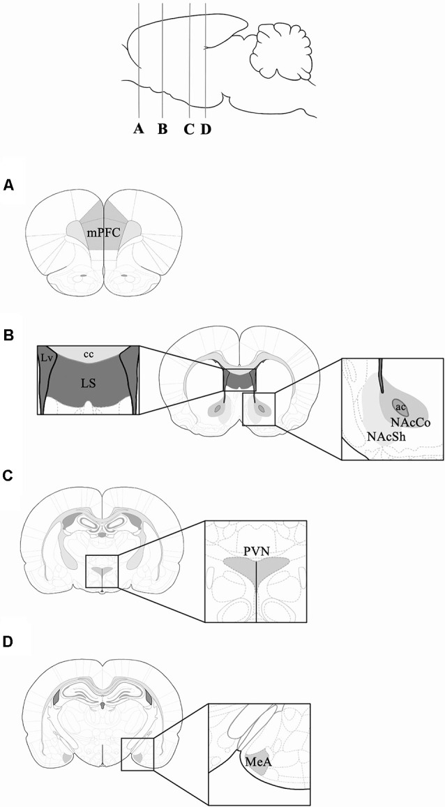
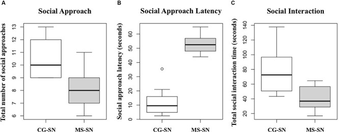
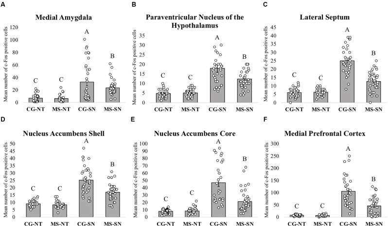
Maternal separation has been shown to disrupt proper brain development and maturation, having profound consequences on the neuroendocrine systems in charge of the stress response, and has been shown to induce behavioral and cognitive abnormalities. At the behavioral level, maternal separation has been shown to increase offensive play-fighting in juvenile individuals and reduce social interest in adulthood. Since most of the studies that have evaluated the consequences of maternal separation on social behavior have focused on behavioral analysis, there is a need for a further understanding of the neuronal mechanisms underlying the changes in social behavior induced by maternal separation. Therefore, the aim of the present research was to assess the long-term effects of maternal separation on social interaction behavior and to assess the activity of several brain regions involved in the processing of social cues and reward upon social novelty exposure, using c-Fos immunohistochemistry as a marker of neuronal activity. Male Wistar rats were subjected to 4 h maternal separation during the neonatal period, 9:00 h-13:00 h from postnatal day 1 to 21, and exposed to social novelty during adulthood. After social novelty exposure, brains were fixed and coronal sections of the medial amygdala, lateral septum (LS), paraventricular nucleus of the hypothalamus, nucleus accumbens, and medial prefrontal cortex were obtained for c-Fos immunohistochemistry. Maternally separated rats spent less time investigating the novel peer, suggesting that maternal separation reduces social approach motivation. Furthermore, maternal separation reduced the number of c-Fos positive cells of the medial amygdala, paraventricular nucleus of the hypothalamus, LS, nucleus accumbens, and medial prefrontal cortex upon social novelty exposure. These findings suggest that maternal separation can reduce the plastic capacity of several brain nuclei, which constitute a physiological basis for the emergence of behavioral disorders presented later in life reported to be linked to early life adversity.
母婴分离已被证明会扰乱大脑的正常发育和成熟,对负责应激反应的神经内分泌系统产生深远影响,并已被证明会导致行为和认知异常。在行为层面,母婴分离已被证明会增加幼年个体的攻击性打斗行为,并降低成年后的社交兴趣。由于大多数评估母婴分离对社会行为影响的研究都集中在行为分析上,因此有必要进一步了解母婴分离诱导的社会行为变化背后的神经元机制。因此,本研究的目的是评估母婴分离对社会互动行为的长期影响,并使用c-Fos免疫组织化学作为神经元活动的标志物,评估在接触社会新奇事物时参与处理社会线索和奖励的几个脑区的活动。雄性Wistar大鼠在新生期(出生后第1天至21天的9:00至13:00)经历4小时的母婴分离,并在成年期接触社会新奇事物。在接触社会新奇事物后,将大脑固定,并获取内侧杏仁核、外侧隔区(LS)、下丘脑室旁核、伏隔核和内侧前额叶皮质的冠状切片进行c-Fos免疫组织化学分析。母婴分离的大鼠花在探究新奇同伴上的时间更少,这表明母婴分离会降低社会接近动机。此外,母婴分离减少了在接触社会新奇事物时内侧杏仁核、下丘脑室旁核、LS、伏隔核和内侧前额叶皮质中c-Fos阳性细胞的数量。这些发现表明,母婴分离会降低几个脑核的可塑性,这构成了据报道与早期生活逆境相关的后期出现的行为障碍的生理基础。