• 文献检索
  • 文档翻译
  • 深度研究
  • 学术资讯
  • Suppr Zotero 插件Zotero 插件
  • 邀请有礼
  • 套餐&价格
  • 历史记录
应用&插件
Suppr Zotero 插件Zotero 插件浏览器插件Mac 客户端Windows 客户端微信小程序
定价
高级版会员购买积分包购买API积分包
服务
文献检索文档翻译深度研究API 文档MCP 服务
关于我们
关于 Suppr公司介绍联系我们用户协议隐私条款
关注我们

Suppr 超能文献

核心技术专利:CN118964589B侵权必究
粤ICP备2023148730 号-1Suppr @ 2026

文献检索

告别复杂PubMed语法,用中文像聊天一样搜索,搜遍4000万医学文献。AI智能推荐,让科研检索更轻松。

立即免费搜索

文件翻译

保留排版,准确专业,支持PDF/Word/PPT等文件格式,支持 12+语言互译。

免费翻译文档

深度研究

AI帮你快速写综述,25分钟生成高质量综述,智能提取关键信息,辅助科研写作。

立即免费体验

肌球蛋白-V 复合物在三种核苷酸状态下的高分辨率结构为力产生机制提供了深入了解。

High-resolution structures of the actomyosin-V complex in three nucleotide states provide insights into the force generation mechanism.

机构信息

Department of Structural Biochemistry, Max Planck Institute of Molecular Physiology, Dortmund, Germany.

Department of Pharmacology and Therapeutics and the Myology Institute, University of Florida, Gainesville, United States.

出版信息

Elife. 2021 Nov 23;10:e73724. doi: 10.7554/eLife.73724.

DOI:10.7554/eLife.73724
PMID:34812732
原文链接:https://pmc.ncbi.nlm.nih.gov/articles/PMC8735999/
Abstract

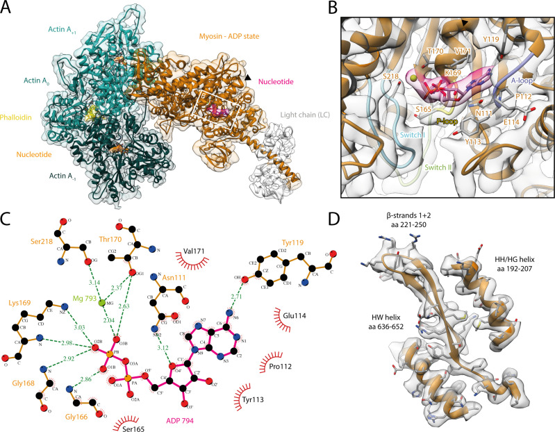
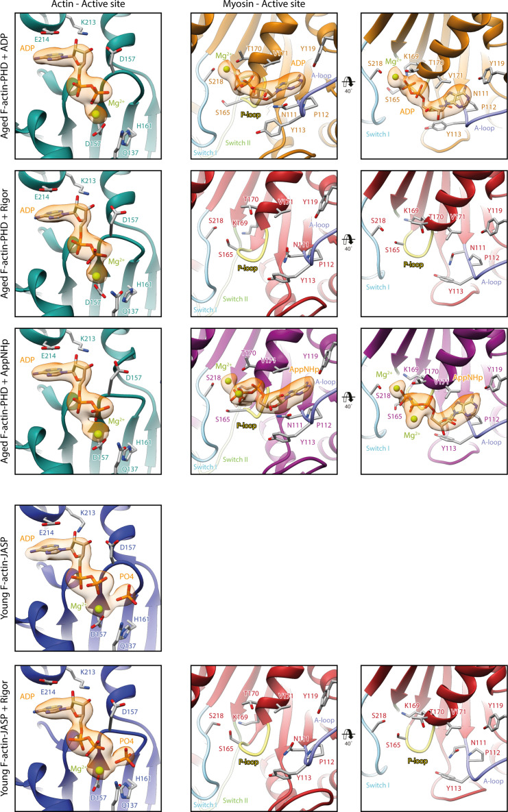
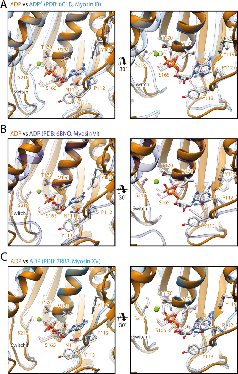
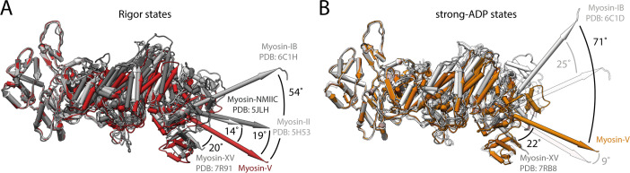
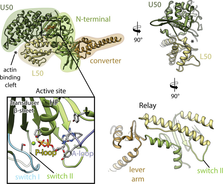
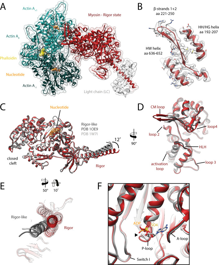
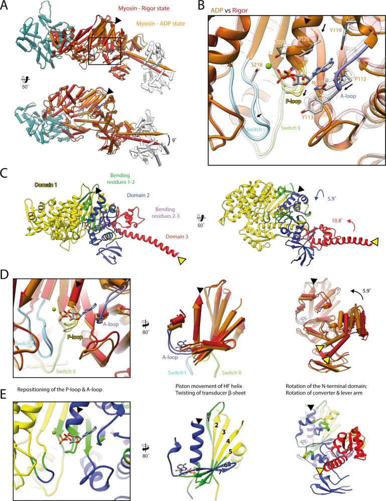
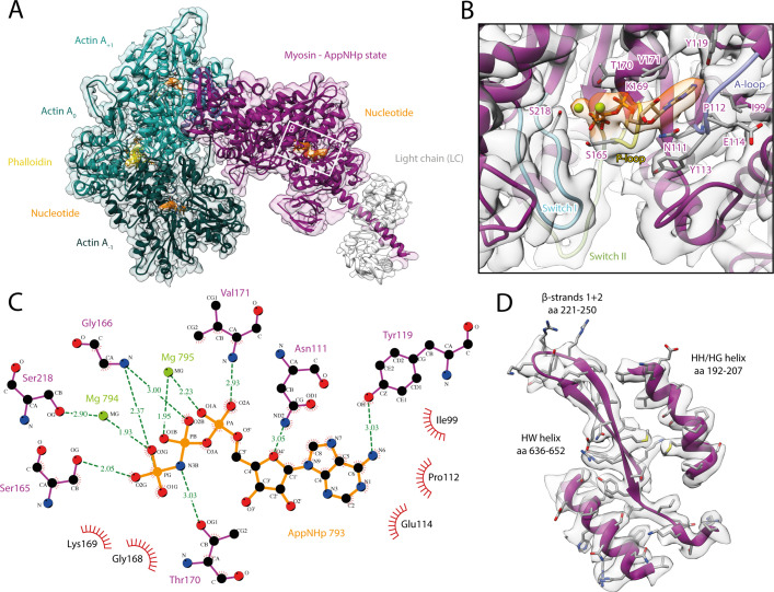
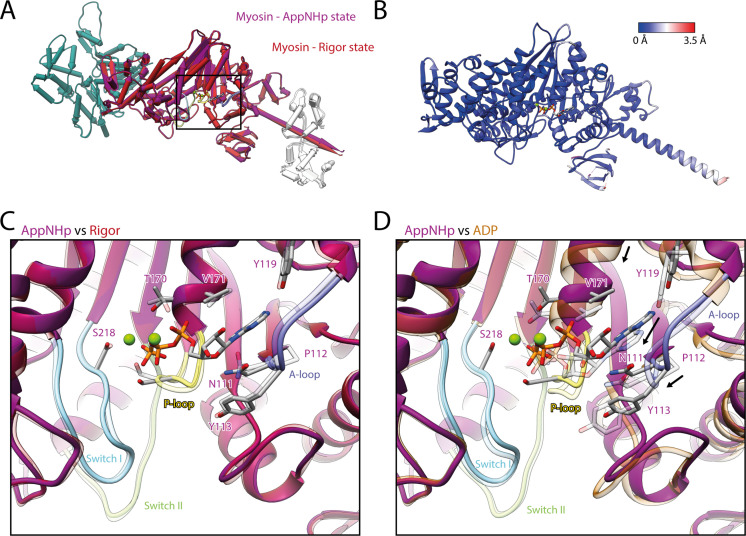
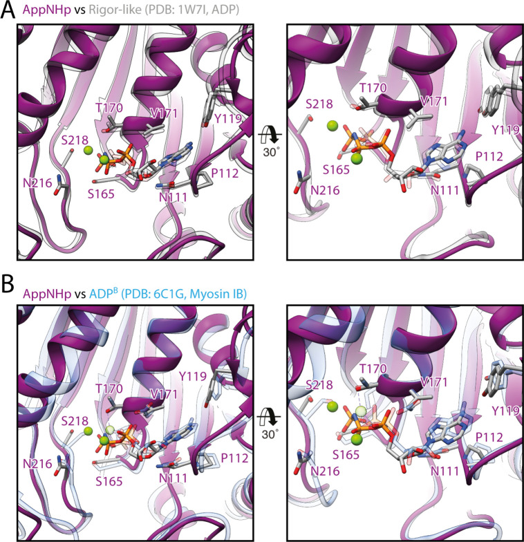
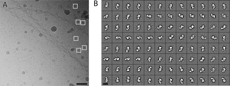
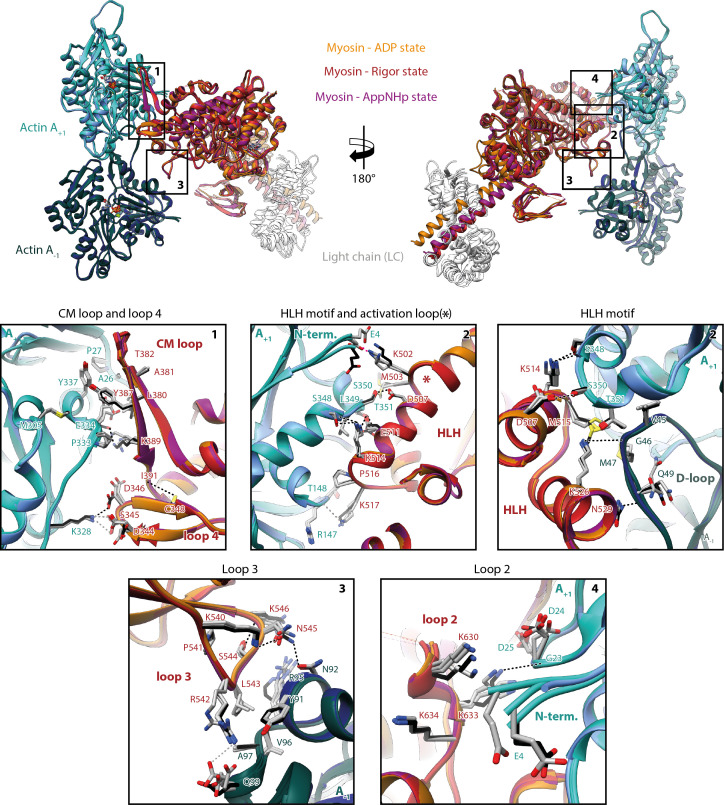
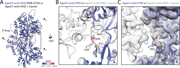
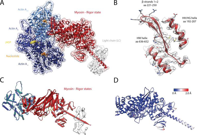
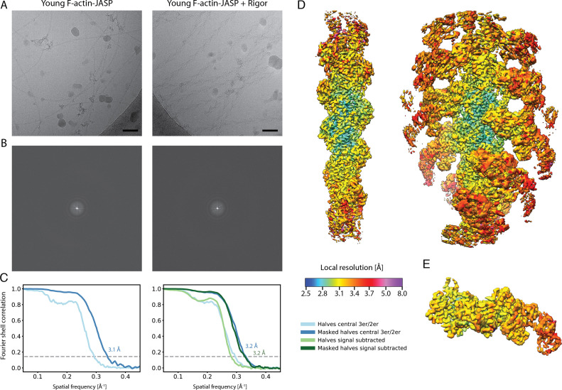
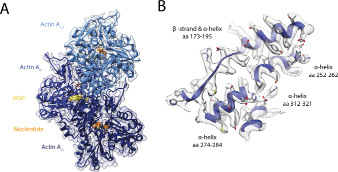
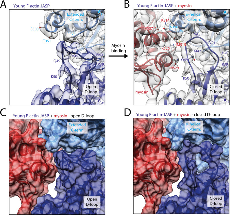
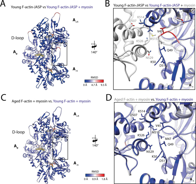
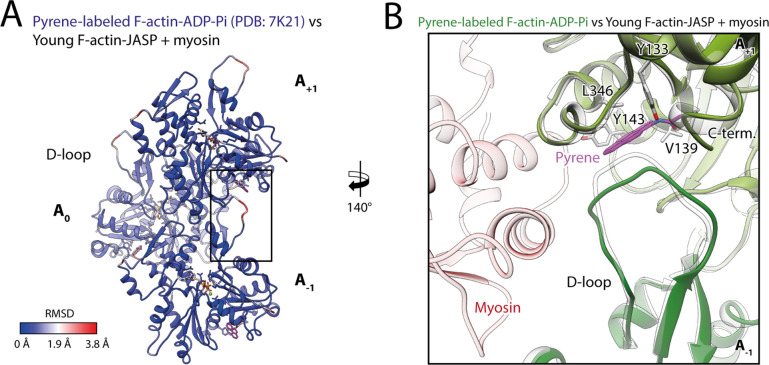
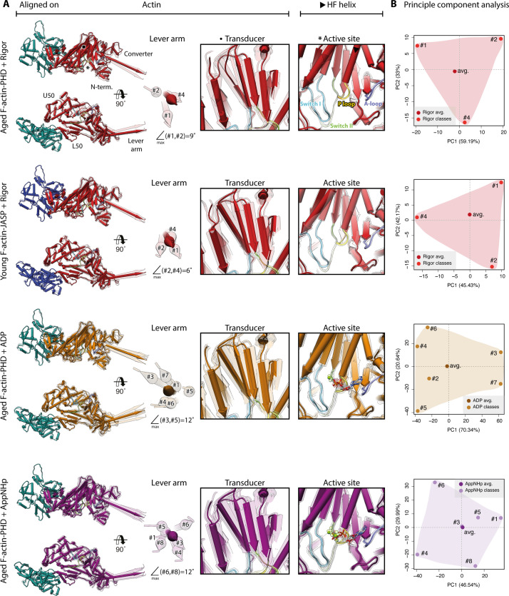
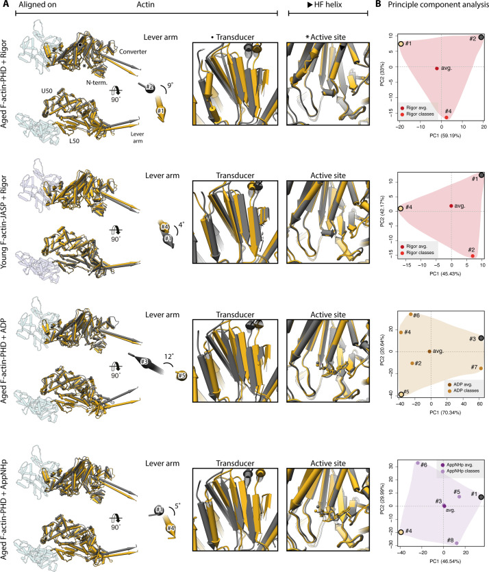
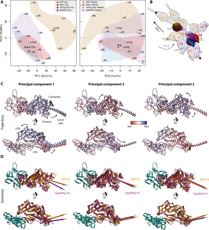
The molecular motor myosin undergoes a series of major structural transitions during its force-producing motor cycle. The underlying mechanism and its coupling to ATP hydrolysis and actin binding are only partially understood, mostly due to sparse structural data on actin-bound states of myosin. Here, we report 26 high-resolution cryo-EM structures of the actomyosin-V complex in the strong-ADP, rigor, and a previously unseen post-rigor transition state that binds the ATP analog AppNHp. The structures reveal a high flexibility of myosin in each state and provide valuable insights into the structural transitions of myosin-V upon ADP release and binding of AppNHp, as well as the actomyosin interface. In addition, they show how myosin is able to specifically alter the structure of F-actin.

摘要

分子马达肌球蛋白在产生力的运动循环中经历一系列主要的结构转变。其潜在机制及其与 ATP 水解和肌动蛋白结合的偶联在很大程度上尚不清楚,主要是因为关于肌球蛋白结合肌动蛋白状态的结构数据稀疏。在这里,我们报告了肌球蛋白 - 肌动球蛋白 V 复合物在强 ADP、僵硬和以前未见的后僵硬过渡状态下的 26 个高分辨率冷冻电镜结构,该复合物结合了 ATP 类似物 AppNHp。这些结构揭示了每种状态下肌球蛋白的高灵活性,并为肌球蛋白 V 在 ADP 释放和 AppNHp 结合以及肌球蛋白 - 肌动蛋白界面时的结构转变提供了有价值的见解。此外,它们还展示了肌球蛋白如何能够特异性地改变 F- 肌动蛋白的结构。

https://cdn.ncbi.nlm.nih.gov/pmc/blobs/baeb/8735999/ca99cfffdcd2/elife-73724-sa2-fig2.jpg
https://cdn.ncbi.nlm.nih.gov/pmc/blobs/baeb/8735999/e53a99c9a521/elife-73724-fig1.jpg
https://cdn.ncbi.nlm.nih.gov/pmc/blobs/baeb/8735999/87228cf7f303/elife-73724-fig1-figsupp1.jpg
https://cdn.ncbi.nlm.nih.gov/pmc/blobs/baeb/8735999/9e2f6a026518/elife-73724-fig1-figsupp2.jpg
https://cdn.ncbi.nlm.nih.gov/pmc/blobs/baeb/8735999/c9cde0cb64bd/elife-73724-fig1-figsupp3.jpg
https://cdn.ncbi.nlm.nih.gov/pmc/blobs/baeb/8735999/b492fd116373/elife-73724-fig1-figsupp4.jpg
https://cdn.ncbi.nlm.nih.gov/pmc/blobs/baeb/8735999/0941f0521899/elife-73724-fig1-figsupp5.jpg
https://cdn.ncbi.nlm.nih.gov/pmc/blobs/baeb/8735999/3ee5c9c6fb5f/elife-73724-fig1-figsupp6.jpg
https://cdn.ncbi.nlm.nih.gov/pmc/blobs/baeb/8735999/d4d56fd2ed73/elife-73724-fig2.jpg
https://cdn.ncbi.nlm.nih.gov/pmc/blobs/baeb/8735999/39ee62788f8c/elife-73724-fig3.jpg
https://cdn.ncbi.nlm.nih.gov/pmc/blobs/baeb/8735999/7ac331e14120/elife-73724-fig4.jpg
https://cdn.ncbi.nlm.nih.gov/pmc/blobs/baeb/8735999/d0669d988e7d/elife-73724-fig4-figsupp1.jpg
https://cdn.ncbi.nlm.nih.gov/pmc/blobs/baeb/8735999/deabf40c6db6/elife-73724-fig4-figsupp2.jpg
https://cdn.ncbi.nlm.nih.gov/pmc/blobs/baeb/8735999/3b7ff91fc90d/elife-73724-fig4-figsupp3.jpg
https://cdn.ncbi.nlm.nih.gov/pmc/blobs/baeb/8735999/090f76fb381d/elife-73724-fig5.jpg
https://cdn.ncbi.nlm.nih.gov/pmc/blobs/baeb/8735999/23275b222524/elife-73724-fig6.jpg
https://cdn.ncbi.nlm.nih.gov/pmc/blobs/baeb/8735999/d1c5cce1eb24/elife-73724-fig7.jpg
https://cdn.ncbi.nlm.nih.gov/pmc/blobs/baeb/8735999/cac406a45255/elife-73724-fig7-figsupp1.jpg
https://cdn.ncbi.nlm.nih.gov/pmc/blobs/baeb/8735999/8efbab65100f/elife-73724-fig7-figsupp2.jpg
https://cdn.ncbi.nlm.nih.gov/pmc/blobs/baeb/8735999/05fb0b334cab/elife-73724-fig8.jpg
https://cdn.ncbi.nlm.nih.gov/pmc/blobs/baeb/8735999/570bd38be2eb/elife-73724-fig8-figsupp1.jpg
https://cdn.ncbi.nlm.nih.gov/pmc/blobs/baeb/8735999/144ee0e53ea3/elife-73724-fig8-figsupp2.jpg
https://cdn.ncbi.nlm.nih.gov/pmc/blobs/baeb/8735999/bea0ac797e99/elife-73724-fig9.jpg
https://cdn.ncbi.nlm.nih.gov/pmc/blobs/baeb/8735999/06d634605138/elife-73724-fig9-figsupp1.jpg
https://cdn.ncbi.nlm.nih.gov/pmc/blobs/baeb/8735999/adfc0a405efa/elife-73724-fig10.jpg
https://cdn.ncbi.nlm.nih.gov/pmc/blobs/baeb/8735999/974d1067a63a/elife-73724-sa2-fig1.jpg
https://cdn.ncbi.nlm.nih.gov/pmc/blobs/baeb/8735999/ca99cfffdcd2/elife-73724-sa2-fig2.jpg

相似文献

1
High-resolution structures of the actomyosin-V complex in three nucleotide states provide insights into the force generation mechanism.肌球蛋白-V 复合物在三种核苷酸状态下的高分辨率结构为力产生机制提供了深入了解。
Elife. 2021 Nov 23;10:e73724. doi: 10.7554/eLife.73724.
2
Cryo-EM structure of a human cytoplasmic actomyosin complex at near-atomic resolution.Cryo-EM 结构解析近原子分辨率的人细胞质肌球蛋白复合物
Nature. 2016 Jun 30;534(7609):724-8. doi: 10.1038/nature18295. Epub 2016 Jun 20.
3
Magnesium, ADP, and actin binding linkage of myosin V: evidence for multiple myosin V-ADP and actomyosin V-ADP states.肌球蛋白V的镁离子、二磷酸腺苷及肌动蛋白结合连接:多种肌球蛋白V-二磷酸腺苷及肌动蛋白-肌球蛋白V-二磷酸腺苷状态的证据
Biochemistry. 2005 Jun 21;44(24):8826-40. doi: 10.1021/bi0473509.
4
High-resolution cryo-EM structures of actin-bound myosin states reveal the mechanism of myosin force sensing.高分辨率冷冻电镜结构的肌球蛋白结合肌动蛋白状态揭示了肌球蛋白力感应的机制。
Proc Natl Acad Sci U S A. 2018 Feb 6;115(6):1292-1297. doi: 10.1073/pnas.1718316115. Epub 2018 Jan 22.
5
Three myosin V structures delineate essential features of chemo-mechanical transduction.三种肌球蛋白V结构描绘了化学机械转导的基本特征。
EMBO J. 2004 Nov 24;23(23):4527-37. doi: 10.1038/sj.emboj.7600458. Epub 2004 Oct 28.
6
Cryo-EM structures reveal specialization at the myosin VI-actin interface and a mechanism of force sensitivity.冷冻电镜结构揭示肌球蛋白 VI-肌动蛋白界面的特化和力敏感性的机制。
Elife. 2017 Dec 4;6:e31125. doi: 10.7554/eLife.31125.
7
High affinity binding of brain myosin-Va to F-actin induced by calcium in the presence of ATP.在ATP存在的情况下,钙诱导脑肌球蛋白-Va与F-肌动蛋白的高亲和力结合。
J Biol Chem. 2001 Oct 26;276(43):39812-8. doi: 10.1074/jbc.M102583200. Epub 2001 Aug 21.
8
A structural state of the myosin V motor without bound nucleotide.肌球蛋白V马达无结合核苷酸时的结构状态。
Nature. 2003 Sep 25;425(6956):419-23. doi: 10.1038/nature01927.
9
The structural basis of myosin V processive movement as revealed by electron cryomicroscopy.冷冻电子显微镜揭示的肌球蛋白V持续运动的结构基础。
Mol Cell. 2005 Sep 2;19(5):595-605. doi: 10.1016/j.molcel.2005.07.015.
10
Allosteric communication in myosin V: from small conformational changes to large directed movements.肌球蛋白V中的变构通讯:从小的构象变化到大的定向运动
PLoS Comput Biol. 2008 Aug 15;4(8):e1000129. doi: 10.1371/journal.pcbi.1000129.

引用本文的文献

1
Swinging lever mechanism of myosin directly shown by time-resolved cryo-EM.通过时间分辨冷冻电镜直接展示的肌球蛋白摆动杠杆机制
Nature. 2025 Apr 9. doi: 10.1038/s41586-025-08876-5.
2
High-resolution structures of Myosin-IC reveal a unique actin-binding orientation, ADP release pathway, and power stroke trajectory.肌球蛋白-IC的高分辨率结构揭示了独特的肌动蛋白结合方向、ADP释放途径和动力冲程轨迹。
Proc Natl Acad Sci U S A. 2025 Mar 4;122(9):e2415457122. doi: 10.1073/pnas.2415457122. Epub 2025 Feb 27.
3
Molecular mechanisms of hotspot variants in cytoskeletal β-actin associated with Baraitser-Winter syndrome.

本文引用的文献

1
Structural basis for tunable control of actin dynamics by myosin-15 in mechanosensory stereocilia.肌球蛋白-15在机械感受性静纤毛中对肌动蛋白动力学进行可调控制的结构基础。
Sci Adv. 2022 Jul 22;8(29):eabl4733. doi: 10.1126/sciadv.abl4733. Epub 2022 Jul 20.
2
High-resolution structures of malaria parasite actomyosin and actin filaments.疟原虫肌球蛋白和肌动蛋白丝的高分辨率结构。
PLoS Pathog. 2022 Apr 4;18(4):e1010408. doi: 10.1371/journal.ppat.1010408. eCollection 2022 Apr.
3
The actomyosin interface contains an evolutionary conserved core and an ancillary interface involved in specificity.
与巴赖特-温特综合征相关的细胞骨架β-肌动蛋白热点变异的分子机制
FEBS J. 2025 Sep;292(18):4898-4917. doi: 10.1111/febs.70018. Epub 2025 Feb 10.
4
High resolution structures of Myosin-IC reveal a unique actin-binding orientation, ADP release pathway, and power stroke trajectory.肌球蛋白-IC的高分辨率结构揭示了独特的肌动蛋白结合方向、ADP释放途径和动力冲程轨迹。
bioRxiv. 2025 Jan 30:2025.01.10.632429. doi: 10.1101/2025.01.10.632429.
5
Myosin forces elicit an F-actin structural landscape that mediates mechanosensitive protein recognition.肌球蛋白力引发一种F-肌动蛋白结构格局,该格局介导机械敏感蛋白识别。
bioRxiv. 2024 Aug 17:2024.08.15.608188. doi: 10.1101/2024.08.15.608188.
6
Insights into Actin Isoform-Specific Interactions with Myosin via Computational Analysis.通过计算分析深入了解肌球蛋白与肌动蛋白同工型的特异性相互作用。
Molecules. 2024 Jun 23;29(13):2992. doi: 10.3390/molecules29132992.
7
Switch-2 determines MgADP-release kinetics and fine-tunes the duty ratio of class-1 myosins.Switch-2决定MgADP释放动力学并微调1类肌球蛋白的占空比。
Front Physiol. 2024 Jun 3;15:1393952. doi: 10.3389/fphys.2024.1393952. eCollection 2024.
8
Myosin's powerstroke transitions define atomic scale movement of cardiac thin filament tropomyosin.肌球蛋白的力循环转变定义了心肌细肌丝原肌球蛋白的原子尺度运动。
J Gen Physiol. 2024 May 6;156(5). doi: 10.1085/jgp.202413538. Epub 2024 Apr 12.
9
Sequence Alignment-Based Prediction of Myosin 7A: Structural Implications and Protein Interactions.基于序列比对的肌球蛋白 7A 预测:结构影响与蛋白相互作用。
Int J Mol Sci. 2024 Mar 16;25(6):3365. doi: 10.3390/ijms25063365.
10
Myosin-5 varies its step length to carry cargo straight along the irregular F-actin track.肌球蛋白-5 改变其步长,以沿着不规则的 F-肌动蛋白轨道直接携带货物。
Proc Natl Acad Sci U S A. 2024 Mar 26;121(13):e2401625121. doi: 10.1073/pnas.2401625121. Epub 2024 Mar 20.
肌动球蛋白界面包含一个进化上保守的核心和一个参与特异性的辅助界面。
Nat Commun. 2021 Mar 25;12(1):1892. doi: 10.1038/s41467-021-22093-4.
4
CryoDRGN: reconstruction of heterogeneous cryo-EM structures using neural networks.CryoDRGN:使用神经网络重建异质冷冻电镜结构。
Nat Methods. 2021 Feb;18(2):176-185. doi: 10.1038/s41592-020-01049-4. Epub 2021 Feb 4.
5
Cryo-EM Resolves Molecular Recognition Of An Optojasp Photoswitch Bound To Actin Filaments In Both Switch States.低温电子显微镜解析光控开关分子与肌动蛋白丝结合的分子识别,该光控开关在两种开关状态下都被结合。
Angew Chem Int Ed Engl. 2021 Apr 12;60(16):8678-8682. doi: 10.1002/anie.202013193. Epub 2021 Mar 4.
6
Cryo-electron microscopy structures of pyrene-labeled ADP-P- and ADP-actin filaments.芘标记的 ADP-P-和 ADP-肌动蛋白纤维的冷冻电子显微镜结构。
Nat Commun. 2020 Nov 19;11(1):5897. doi: 10.1038/s41467-020-19762-1.
7
TranSPHIRE: automated and feedback-optimized on-the-fly processing for cryo-EM.TranSPHIRE:用于冷冻电镜的自动化和反馈优化的实时处理。
Nat Commun. 2020 Nov 11;11(1):5716. doi: 10.1038/s41467-020-19513-2.
8
High-Resolution Cryo-EM Structure of the Cardiac Actomyosin Complex.心脏肌球蛋白复合物的高分辨率冷冻电镜结构。
Structure. 2021 Jan 7;29(1):50-60.e4. doi: 10.1016/j.str.2020.09.013. Epub 2020 Oct 15.
9
Alternative N-terminal regions of myosin heavy chain II regulate communication of the purine binding loop with the essential light chain.肌球蛋白重链 II 的替代 N 端区域调节嘌呤结合环与必需轻链的通讯。
J Biol Chem. 2020 Oct 16;295(42):14522-14535. doi: 10.1074/jbc.RA120.014684. Epub 2020 Aug 19.
10
Cryo-EM and Molecular Docking Shows Myosin Loop 4 Contacts Actin and Tropomyosin on Thin Filaments.低温电子显微镜和分子对接显示肌球蛋白环 4 与细肌丝上的肌动蛋白和原肌球蛋白接触。
Biophys J. 2020 Aug 18;119(4):821-830. doi: 10.1016/j.bpj.2020.07.006. Epub 2020 Jul 16.