Carl Ayala G, Reynolds Matthew J, Gurel Pinar S, Phua Donovan Y Z, Sun Xiaoyu, Mei Lin, Hamilton Keith, Takagi Yasuharu, Noble Alex J, Sellers James R, Alushin Gregory M
Laboratory of Structural Biophysics and Mechanobiology, The Rockefeller University, New York, NY, USA.
Tri-Institutional Program in Chemical Biology, The Rockefeller University, New York, NY, USA.
bioRxiv. 2024 Aug 17:2024.08.15.608188. doi: 10.1101/2024.08.15.608188.
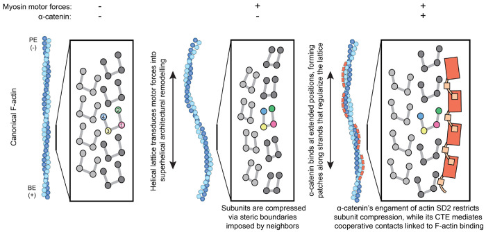
Cells mechanically interface with their surroundings through cytoskeleton-linked adhesions, allowing them to sense physical cues that instruct development and drive diseases such as cancer. Contractile forces generated by myosin motor proteins mediate these mechanical signal transduction processes through unclear protein structural mechanisms. Here, we show that myosin forces elicit structural changes in actin filaments (F-actin) that modulate binding by the mechanosensitive adhesion protein α-catenin. Using correlative cryo-fluorescence microscopy and cryo-electron tomography, we identify F-actin featuring domains of nanoscale oscillating curvature at cytoskeleton-adhesion interfaces enriched in zyxin, a marker of actin-myosin generated traction forces. We next introduce a reconstitution system for visualizing F-actin in the presence of myosin forces with cryo-electron microscopy, which reveals morphologically similar superhelical F-actin spirals. In simulations, transient forces mimicking tugging and release of filaments by motors produce spirals, supporting a mechanistic link to myosin's ATPase mechanochemical cycle. Three-dimensional reconstruction of spirals uncovers extensive asymmetric remodeling of F-actin's helical lattice. This is recognized by α-catenin, which cooperatively binds along individual strands, preferentially engaging interfaces featuring extended inter-subunit distances while simultaneously suppressing rotational deviations to regularize the lattice. Collectively, we find that myosin forces can deform F-actin, generating a conformational landscape that is detected and reciprocally modulated by a mechanosensitive protein, providing a direct structural glimpse at active force transduction through the cytoskeleton.
细胞通过细胞骨架连接的黏附与周围环境进行机械接触,使它们能够感知指导发育并引发癌症等疾病的物理线索。肌球蛋白运动蛋白产生的收缩力通过尚不清楚的蛋白质结构机制介导这些机械信号转导过程。在这里,我们表明肌球蛋白力会引起肌动蛋白丝(F-肌动蛋白)的结构变化,从而调节机械敏感黏附蛋白α-连环蛋白的结合。通过相关冷冻荧光显微镜和冷冻电子断层扫描,我们在富含桩蛋白(肌动蛋白-肌球蛋白产生的牵引力的标志物)的细胞骨架-黏附界面处鉴定出具有纳米级振荡曲率结构域的F-肌动蛋白。接下来,我们引入了一个重组系统,用于在存在肌球蛋白力的情况下用冷冻电子显微镜观察F-肌动蛋白,该系统揭示了形态上相似的超螺旋F-肌动蛋白螺旋。在模拟中,模仿马达对细丝的牵拉和释放的瞬态力产生螺旋,支持了与肌球蛋白ATP酶机械化学循环的机械联系。螺旋的三维重建揭示了F-肌动蛋白螺旋晶格的广泛不对称重塑。这被α-连环蛋白识别,α-连环蛋白沿着单链协同结合,优先与具有延长的亚基间距离的界面结合,同时抑制旋转偏差以规范晶格。总体而言,我们发现肌球蛋白力可以使F-肌动蛋白变形,产生一种构象景观,该景观被一种机械敏感蛋白检测并相互调节,为通过细胞骨架的主动力转导提供了直接的结构视角。