• 文献检索
  • 文档翻译
  • 深度研究
  • 学术资讯
  • Suppr Zotero 插件Zotero 插件
  • 邀请有礼
  • 套餐&价格
  • 历史记录
应用&插件
Suppr Zotero 插件Zotero 插件浏览器插件Mac 客户端Windows 客户端微信小程序
定价
高级版会员购买积分包购买API积分包
服务
文献检索文档翻译深度研究API 文档MCP 服务
关于我们
关于 Suppr公司介绍联系我们用户协议隐私条款
关注我们

Suppr 超能文献

核心技术专利:CN118964589B侵权必究
粤ICP备2023148730 号-1Suppr @ 2026

文献检索

告别复杂PubMed语法,用中文像聊天一样搜索,搜遍4000万医学文献。AI智能推荐,让科研检索更轻松。

立即免费搜索

文件翻译

保留排版,准确专业,支持PDF/Word/PPT等文件格式,支持 12+语言互译。

免费翻译文档

深度研究

AI帮你快速写综述,25分钟生成高质量综述,智能提取关键信息,辅助科研写作。

立即免费体验

临床表型调节颞叶癫痫患者大脑的髓鞘和铁含量。

Clinical phenotype modulates brain's myelin and iron content in temporal lobe epilepsy.

机构信息

LREN, Centre for Research in Neuroscience, Department of Clinical Neurosciences, Lausanne University Hospital and University of Lausanne, Mont Paisible 16, 1011, Lausanne, Switzerland.

EEG and Epilepsy Unit, Department of Neurology, Department of Clinical Neurosciences, University Hospitals and Faculty of Medicine Geneva, Geneva, Switzerland.

出版信息

Brain Struct Funct. 2022 Apr;227(3):901-911. doi: 10.1007/s00429-021-02428-z. Epub 2021 Nov 24.

DOI:10.1007/s00429-021-02428-z
PMID:34817680
原文链接:https://pmc.ncbi.nlm.nih.gov/articles/PMC8930791/
Abstract

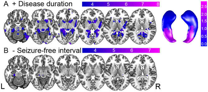
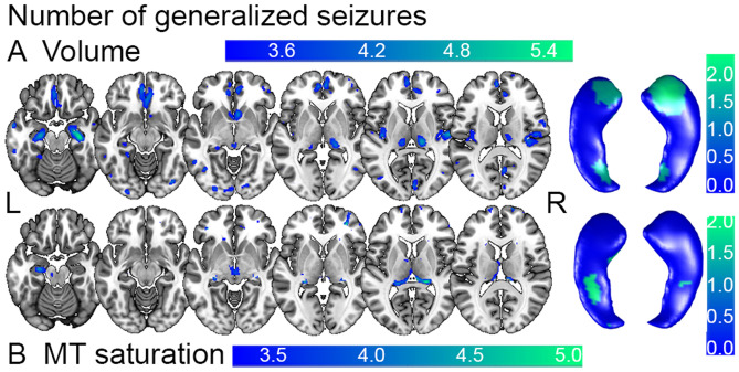
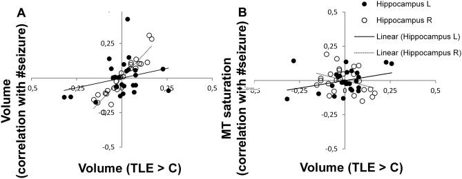
Temporal lobe epilepsy (TLE) is associated with brain pathology extending beyond temporal lobe structures. We sought to look for informative patterns of brain tissue properties in TLE that go beyond the established morphometry differences. We hypothesised that volume differences, particularly in hippocampus, will be paralleled by changes in brain microstructure. The cross-sectional study included TLE patients (n = 25) from a primary care center and sex-/age-matched healthy controls (n = 55). We acquired quantitative relaxometry-based magnetic resonance imaging (MRI) data yielding whole-brain maps of grey matter volume, magnetization transfer (MT) saturation, and effective transverse relaxation rate R2* indicative for brain tissue myelin and iron content. For statistical analysis, we used the computational anatomy framework of voxel-based morphometry and voxel-based quantification. There was a positive correlation between seizure activity and MT saturation measures in the ipsilateral hippocampus, paralleled by volume differences bilaterally. Disease duration correlated positively with iron content in the mesial temporal lobe, while seizure freedom was associated with a decrease of iron in the very same region. Our findings demonstrate the link between TLE clinical phenotype and brain anatomy beyond morphometry differences to show the impact of disease burden on specific tissue properties. We provide direct evidence for the differential effect of clinical phenotype characteristics on processes involving tissue myelin and iron in mesial temporal lobe structures. This study offers a proof-of-concept for the investigation of novel imaging biomarkers in focal epilepsy.

摘要

颞叶癫痫(TLE)与超出颞叶结构的脑病理学有关。我们试图寻找 TLE 中超出既定形态测量差异的有意义的脑组织特性模式。我们假设,体积差异,特别是海马体的体积差异,将与脑微观结构的变化相平行。这项横断面研究包括来自初级保健中心的 TLE 患者(n=25)和性别/年龄匹配的健康对照组(n=55)。我们获得了基于定量弛豫率的磁共振成像(MRI)数据,生成了全脑灰质体积、磁化传递(MT)饱和度和有效横向弛豫率 R2*的图谱,这些指标分别代表脑组织结构的髓鞘和铁含量。为了进行统计分析,我们使用了基于体素的形态学和基于体素的定量计算解剖框架。在同侧海马体中,癫痫活动与 MT 饱和度测量呈正相关,双侧海马体体积存在差异。疾病持续时间与内侧颞叶的铁含量呈正相关,而无癫痫发作与同一区域的铁含量减少相关。我们的发现表明,TLE 临床表型与脑解剖之间存在联系,超出了形态测量差异,以显示疾病负担对特定组织特性的影响。我们提供了直接证据,证明临床表型特征对涉及内侧颞叶结构中组织髓鞘和铁的过程有不同的影响。这项研究为局灶性癫痫的新型成像生物标志物的研究提供了概念验证。

https://cdn.ncbi.nlm.nih.gov/pmc/blobs/0c3b/8930791/e72b95905ccd/429_2021_2428_Fig4_HTML.jpg
https://cdn.ncbi.nlm.nih.gov/pmc/blobs/0c3b/8930791/404750719434/429_2021_2428_Fig1_HTML.jpg
https://cdn.ncbi.nlm.nih.gov/pmc/blobs/0c3b/8930791/638dafc38682/429_2021_2428_Fig2_HTML.jpg
https://cdn.ncbi.nlm.nih.gov/pmc/blobs/0c3b/8930791/f0bf6b63b809/429_2021_2428_Fig3_HTML.jpg
https://cdn.ncbi.nlm.nih.gov/pmc/blobs/0c3b/8930791/e72b95905ccd/429_2021_2428_Fig4_HTML.jpg

相似文献

1
Clinical phenotype modulates brain's myelin and iron content in temporal lobe epilepsy.临床表型调节颞叶癫痫患者大脑的髓鞘和铁含量。
Brain Struct Funct. 2022 Apr;227(3):901-911. doi: 10.1007/s00429-021-02428-z. Epub 2021 Nov 24.
2
Trajectories of brain remodeling in temporal lobe epilepsy.颞叶癫痫中的脑重塑轨迹。
J Neurol. 2019 Dec;266(12):3150-3159. doi: 10.1007/s00415-019-09546-z. Epub 2019 Sep 23.
3
Temporal and extratemporal atrophic manifestation of temporal lobe epilepsy using voxel-based morphometry and corticometry: clinical application in lateralization of epileptogenic zone.基于体素形态计量学和皮质测量的颞叶癫痫的时间和颞外萎缩表现:在致痫区定位中的临床应用。
Neurol Sci. 2021 Aug;42(8):3305-3325. doi: 10.1007/s10072-020-05003-2. Epub 2021 Jan 3.
4
Remodeling of brain morphology in temporal lobe epilepsy.颞叶癫痫患者的脑形态重塑。
Brain Behav. 2020 Nov;10(11):e01825. doi: 10.1002/brb3.1825. Epub 2020 Sep 17.
5
Whole-brain voxel-based statistical analysis of gray matter and white matter in temporal lobe epilepsy.颞叶癫痫患者灰质和白质的基于全脑体素的统计分析。
Neuroimage. 2004 Oct;23(2):717-23. doi: 10.1016/j.neuroimage.2004.06.015.
6
Abnormal neurite density and orientation dispersion in unilateral temporal lobe epilepsy detected by advanced diffusion imaging.异常的神经突密度和单侧颞叶癫痫的取向分散通过先进的扩散成像检测到。
Neuroimage Clin. 2018;20:772-782. doi: 10.1016/j.nicl.2018.09.017. Epub 2018 Sep 23.
7
Microstructural imaging in temporal lobe epilepsy: Diffusion imaging changes relate to reduced neurite density.颞叶癫痫的微观结构成像:弥散成像的变化与神经丝密度降低有关。
Neuroimage Clin. 2020;26:102231. doi: 10.1016/j.nicl.2020.102231. Epub 2020 Feb 28.
8
Cerebrocerebellar structural covariance in temporal lobe epilepsy with hippocampal sclerosis.颞叶癫痫伴海马硬化的脑 cerebellum 结构协方差。
Epilepsy Behav. 2020 Oct;111:107180. doi: 10.1016/j.yebeh.2020.107180. Epub 2020 Jun 26.
9
Voxel-based T2 relaxation rate measurements in temporal lobe epilepsy (TLE) with and without mesial temporal sclerosis.基于体素的颞叶癫痫伴或不伴内侧颞叶硬化的T2弛豫率测量
Epilepsia. 2007 Feb;48(2):220-8. doi: 10.1111/j.1528-1167.2006.00916.x.
10
Auras and clinical features in temporal lobe epilepsy: a new approach on the basis of voxel-based morphometry.基于体素形态计量学的颞叶癫痫的先兆和临床特征:一种新方法。
Epilepsy Res. 2010 May;89(2-3):327-38. doi: 10.1016/j.eplepsyres.2010.02.006. Epub 2010 Mar 12.

引用本文的文献

1
Inhibiting Inducible Nitric Oxide Synthase with 1400W Reduces Soman (GD)-Induced Ferroptosis in Long-Term Epilepsy-Associated Neuropathology: Structural and Functional Magnetic Resonance Imaging Correlations with Neurobehavior and Brain Pathology.用 1400W 抑制诱导型一氧化氮合酶可减少梭曼(GD)诱导的长期癫痫相关神经病理学中的铁死亡:与神经行为和脑病理学的结构和功能磁共振成像相关性。
J Pharmacol Exp Ther. 2024 Jan 17;388(2):724-738. doi: 10.1124/jpet.123.001929.
2
Identification of ferroptosis-related genes in acute phase of temporal lobe epilepsy based on bioinformatic analysis.基于生物信息学分析鉴定颞叶癫痫急性期的铁死亡相关基因。
BMC Genomics. 2023 Nov 9;24(1):675. doi: 10.1186/s12864-023-09782-8.
3

本文引用的文献

1
Converging patterns of aging-associated brain volume loss and tissue microstructure differences.衰老相关的大脑体积损失和组织微观结构差异的趋同模式。
Neurobiol Aging. 2020 Apr;88:108-118. doi: 10.1016/j.neurobiolaging.2020.01.006. Epub 2020 Jan 15.
2
Apparent thinning of human visual cortex during childhood is associated with myelination.儿童时期人类视觉皮层的明显变薄与髓鞘形成有关。
Proc Natl Acad Sci U S A. 2019 Oct 8;116(41):20750-20759. doi: 10.1073/pnas.1904931116. Epub 2019 Sep 23.
3
Inhibition of ferroptosis processes ameliorates cognitive impairment in kainic acid-induced temporal lobe epilepsy in rats.
Sex-based structural and functional MRI outcomes in the rat brain after soman (GD) exposure-induced status epilepticus.
遭受梭曼(GD)暴露诱导的癫痫持续状态后大鼠脑的基于性别的结构和功能 MRI 结果。
Epilepsia Open. 2023 Jun;8(2):399-410. doi: 10.1002/epi4.12701. Epub 2023 Feb 6.
抑制铁死亡过程可改善海藻酸诱导的大鼠颞叶癫痫中的认知障碍。
Am J Transl Res. 2019 Feb 15;11(2):875-884. eCollection 2019.
4
Preferential susceptibility of limbic cortices to microstructural damage in temporal lobe epilepsy: A quantitative T1 mapping study.颞叶癫痫患者边缘皮质对结构损伤的易感性:一项定量 T1 映射研究。
Neuroimage. 2018 Nov 15;182:294-303. doi: 10.1016/j.neuroimage.2017.06.002. Epub 2017 Jun 3.
5
Nuclei-specific deposits of iron and calcium in the rat thalamus after status epilepticus revealed with quantitative susceptibility mapping (QSM).癫痫持续状态后大鼠丘脑内铁和钙的核特异性沉积的定量磁化率映射(QSM)显示。
J Magn Reson Imaging. 2018 Feb;47(2):554-564. doi: 10.1002/jmri.25777. Epub 2017 Jun 5.
6
Electroconvulsive stimulation results in long-term survival of newly generated hippocampal neurons in rats.电惊厥刺激可使大鼠新生海马神经元长期存活。
Hippocampus. 2017 Jan;27(1):52-60. doi: 10.1002/hipo.22670. Epub 2016 Oct 31.
7
Neurobiological origin of spurious brain morphological changes: A quantitative MRI study.假性脑形态学改变的神经生物学起源:一项定量磁共振成像研究。
Hum Brain Mapp. 2016 May;37(5):1801-15. doi: 10.1002/hbm.23137. Epub 2016 Feb 15.
8
New tissue priors for improved automated classification of subcortical brain structures on MRI.用于改进MRI上皮质下脑结构自动分类的新组织先验信息。
Neuroimage. 2016 Apr 15;130:157-166. doi: 10.1016/j.neuroimage.2016.01.062. Epub 2016 Feb 5.
9
Alterations in hippocampal myelin and oligodendrocyte precursor cells during epileptogenesis.癫痫发生过程中海马髓鞘和少突胶质前体细胞的改变。
Brain Res. 2015 Nov 19;1627:154-64. doi: 10.1016/j.brainres.2015.09.027. Epub 2015 Oct 4.
10
Neuronal hyperactivity accelerates depletion of neural stem cells and impairs hippocampal neurogenesis.神经元活动亢进会加速神经干细胞的消耗,并损害海马体神经发生。
Cell Stem Cell. 2015 May 7;16(5):488-503. doi: 10.1016/j.stem.2015.04.003.