Department of Comparative Medicine, Stanford University, Palo Alto, CA 94305-5410
Department of Comparative Medicine, Stanford University, Palo Alto, CA 94305-5410.
eNeuro. 2021 Dec 27;8(6). doi: 10.1523/ENEURO.0299-21.2021. Print 2021 Nov-Dec.
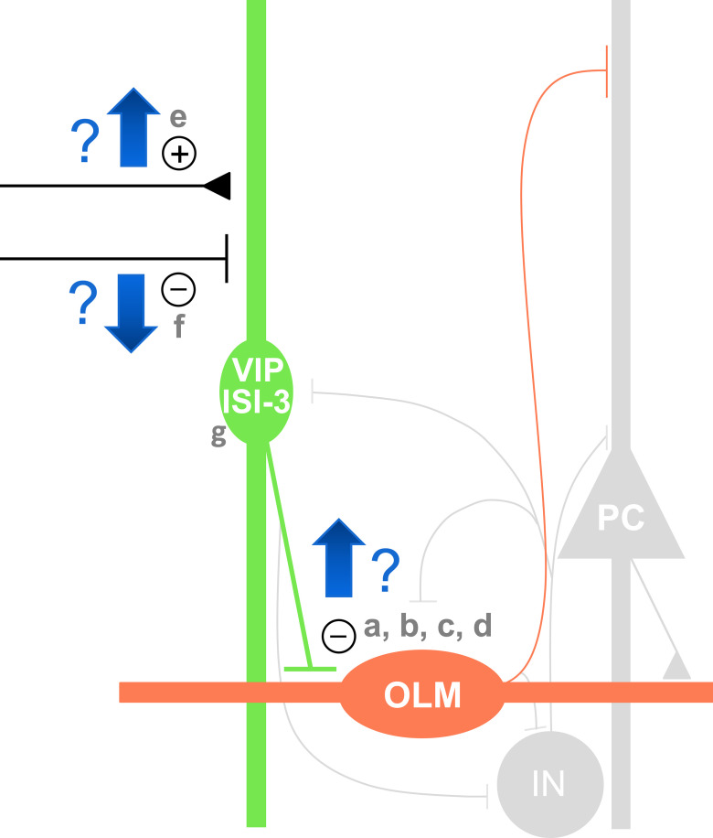
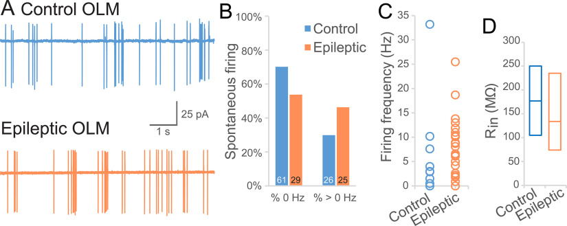
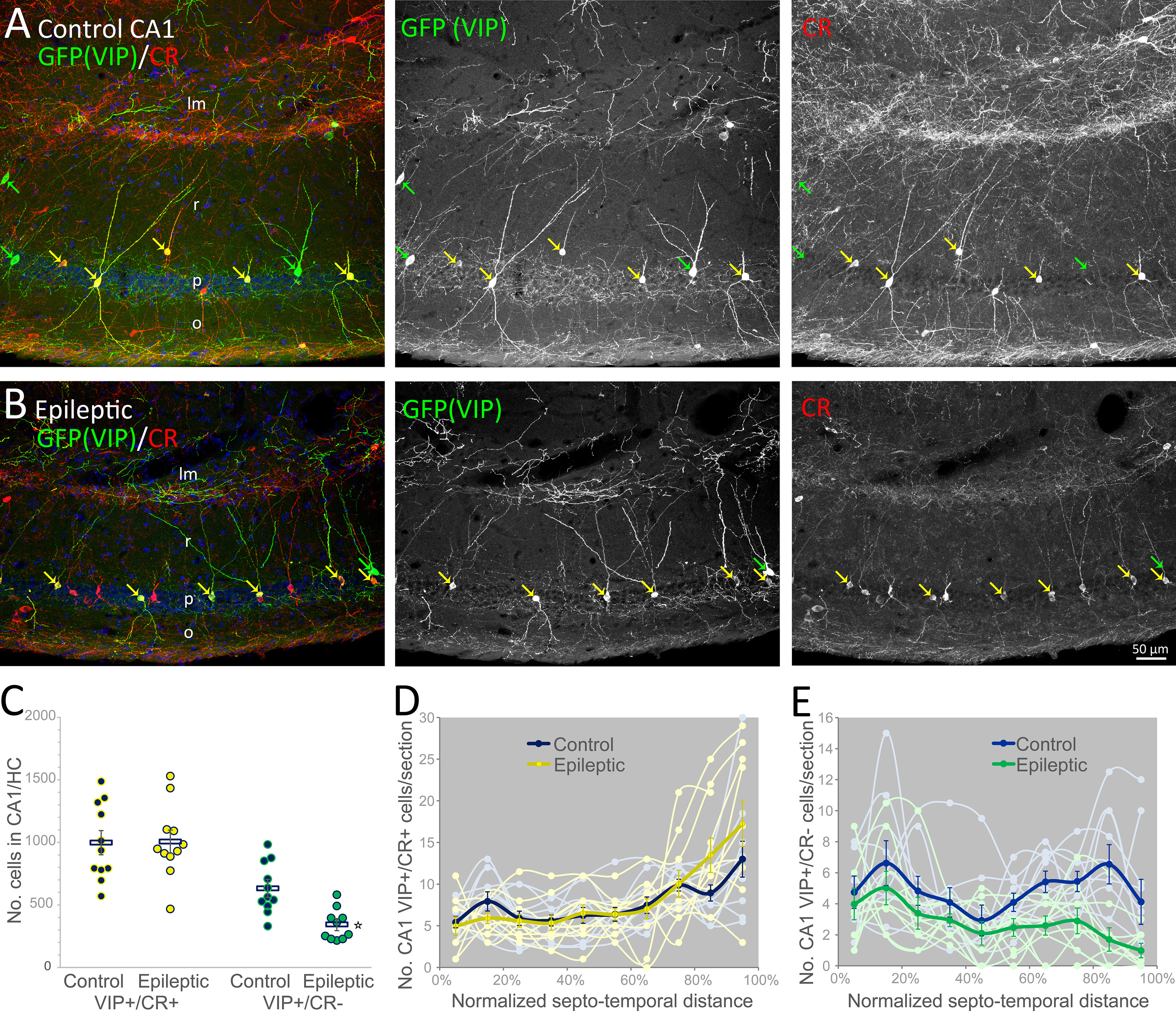
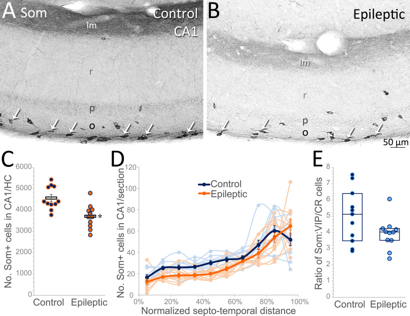
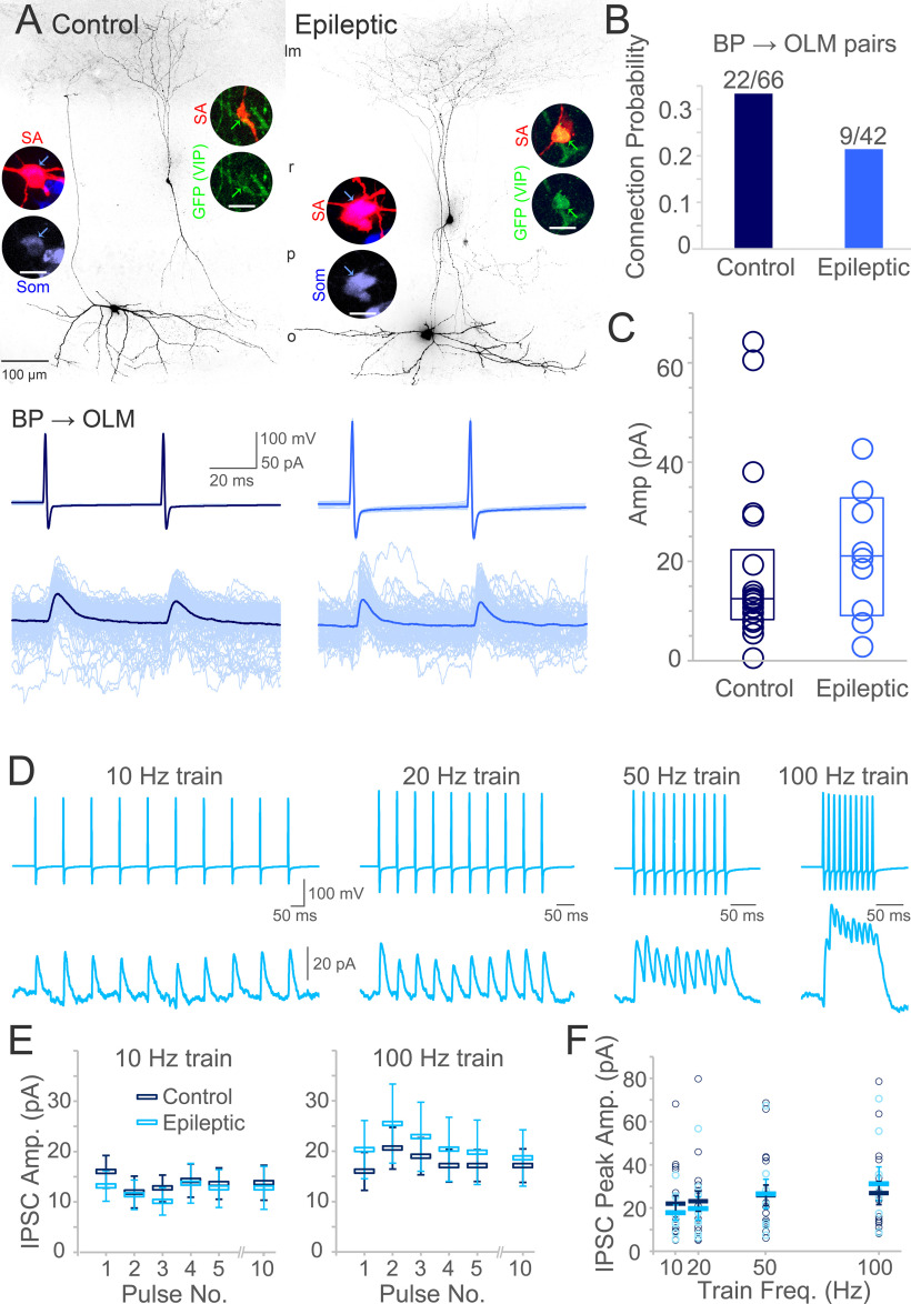
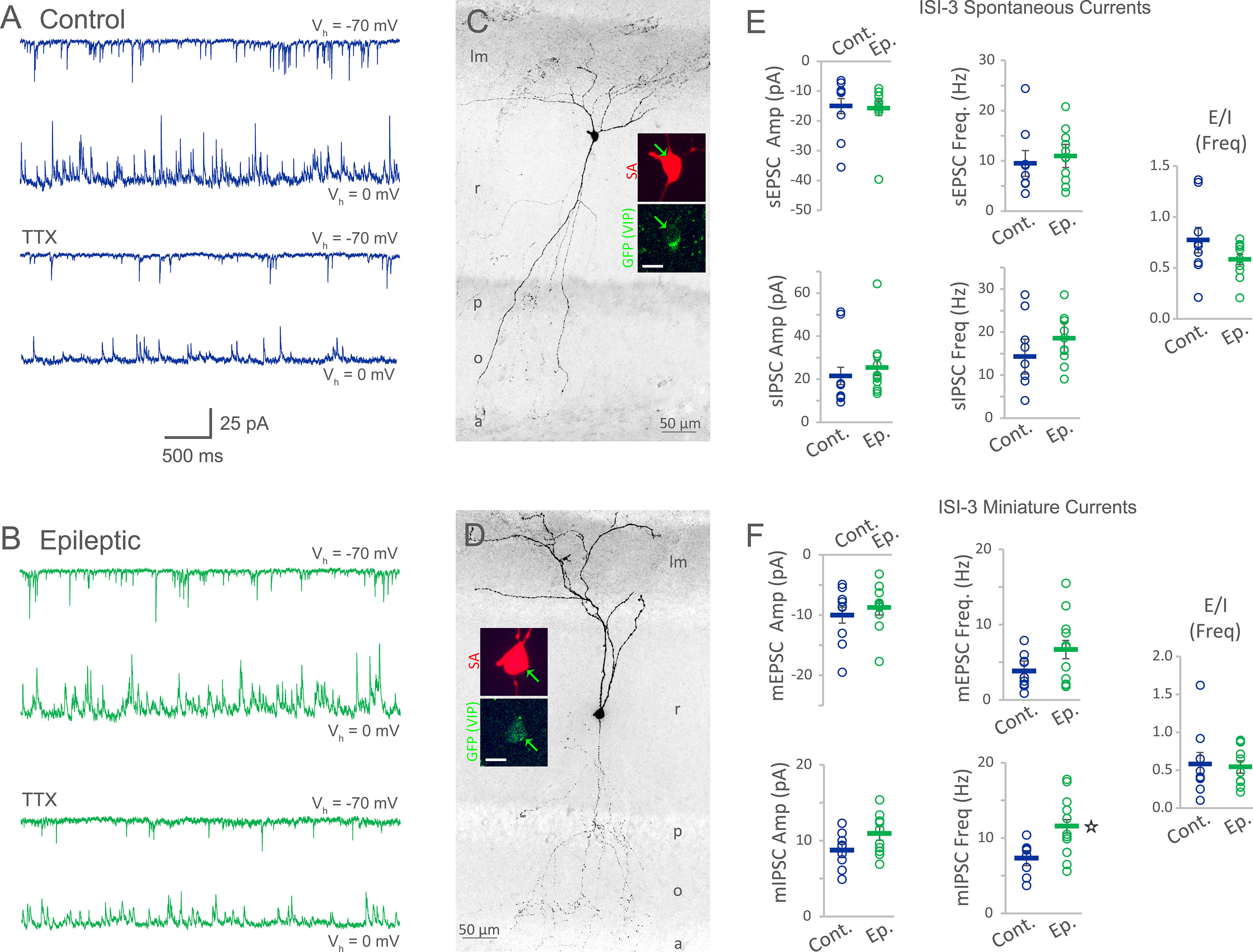
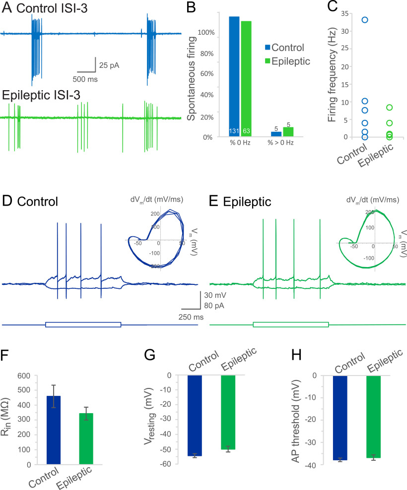
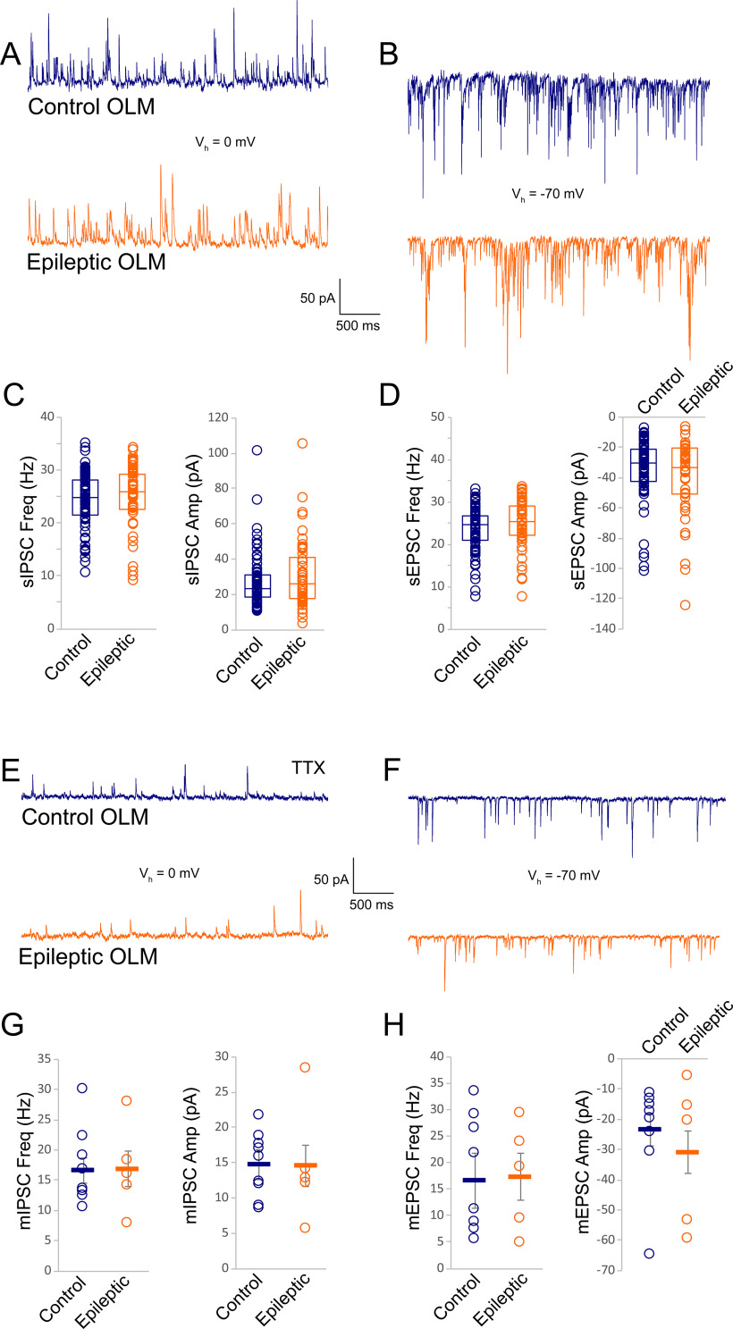
Temporal lobe epilepsy remains a common disorder with no cure and inadequate treatments, potentially because of an incomplete understanding of how seizures start. CA1 pyramidal cells and many inhibitory interneurons increase their firing rate in the seconds-minutes before a spontaneous seizure in epileptic rats. However, some interneurons fail to do so, including those identified as putative interneurons with somata in oriens and axons targeting lacunosum-moleculare (OLM cells). Somatostatin-containing cells, including OLM cells, are the primary target of inhibitory vasoactive intestinal polypeptide and calretinin-expressing (VIP/CR) bipolar interneuron-selective interneurons, type 3 (ISI-3). The objective of this study was to test the hypothesis that in epilepsy inhibition of OLM cells by ISI-3 is abnormally increased, potentially explaining the failure of OLM recruitment when needed most during the ramp up of activity preceding a seizure. Stereological quantification of VIP/CR cells in a model of temporal lobe epilepsy demonstrated that they survive in epileptic mice, despite a reduction in their somatostatin-expressing (Som) cell targets. Paired recordings of unitary IPSCs (uIPSCs) from ISI-3 to OLM cells did not show increased connection probability or increased connection strength, and failure rate was unchanged. When miniature postsynaptic currents in ISI-3 were compared, only mIPSC frequency was increased in epileptic hippocampi. Nevertheless, spontaneous and miniature postsynaptic potentials were unchanged in OLM cells of epileptic mice. These results are not consistent with the hypothesis of hyperinhibition from VIP/CR bipolar cells impeding recruitment of OLM cells in advance of a seizure.
颞叶癫痫仍然是一种常见的疾病,没有治愈方法,治疗效果也不理想,这可能是因为人们对癫痫发作的起始机制还没有完全了解。在癫痫大鼠自发性癫痫发作前的几秒钟到几分钟内,CA1 锥体神经元和许多抑制性中间神经元的放电频率增加。然而,有些中间神经元并没有这样做,包括那些被认为是具有体细胞核和轴突靶向 lacunosum-moleculare(OLM 细胞)的中间神经元。包含生长抑素的细胞,包括 OLM 细胞,是抑制性血管活性肠肽和钙调蛋白表达(VIP/CR)双极中间神经元选择性中间神经元、类型 3(ISI-3)的主要靶标。本研究的目的是验证这样一个假设,即在癫痫中,ISI-3 对 OLM 细胞的抑制作用异常增加,这可能解释了在癫痫发作前活动逐渐增加期间,OLM 细胞在最需要的时候招募失败的原因。在颞叶癫痫模型中,VIP/CR 细胞的立体定量分析表明,尽管它们的生长抑素表达(Som)细胞靶标减少,但它们在癫痫小鼠中仍然存活。从 ISI-3 到 OLM 细胞的成对记录单位 IPSC(uIPSCs)显示,它们的连接概率或连接强度没有增加,失效率也没有改变。当比较 ISI-3 中的微小突触后电流时,只有癫痫海马中的 mIPSC 频率增加。然而,癫痫小鼠 OLM 细胞中的自发性和微小突触后电位没有变化。这些结果与 VIP/CR 双极细胞的超抑制假说不一致,该假说认为 VIP/CR 双极细胞的超抑制会阻碍癫痫发作前 OLM 细胞的募集。