Cannella Roberto, Dioguardi Burgio Marco, Beaufrère Aurélie, Trapani Loïc, Paradis Valérie, Hobeika Christian, Cauchy Francois, Bouattour Mohamed, Vilgrain Valérie, Sartoris Riccardo, Ronot Maxime
Department of Radiology, Hôpital Beaujon, Clichy, France.
Section of Radiology-BiND, University Hospital 'Paolo Giaccone', Palermo, Italy.
JHEP Rep. 2021 Sep 30;3(6):100380. doi: 10.1016/j.jhepr.2021.100380. eCollection 2021 Dec.
BACKGROUND & AIMS: The histopathological subtypes of hepatocellular carcinoma (HCC) are associated with distinct clinical features and prognoses. This study aims to report Liver Imaging Reporting and Data System (LI-RADS)-defined imaging features of different HCC subtypes in a cohort of resected tumours and to assess the influence of HCC subtypes on computed tomography (CT)/magnetic resonance imaging (MRI) LI-RADS categorisation in the subgroup of high-risk patients.
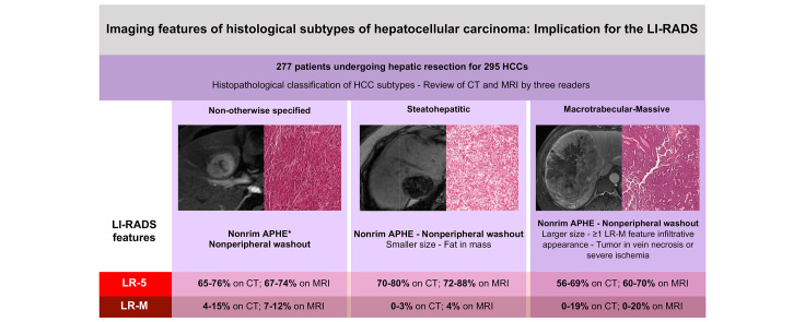
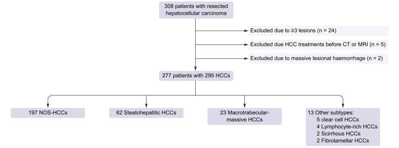
This retrospective institutional review board-approved study included patients with resected HCCs and available histopathological classification. Three radiologists independently reviewed preoperative CT and MRI exams. The readers evaluated the presence of imaging features according to LI-RADS v2018 definitions and provided a LI-RADS category in patients at high risk of HCC. Differences in LI-RADS features and categorisations were assessed for not otherwise specified (NOS-HCC), steatohepatitic (SH-HCC), and macrotrabecular-massive (MTM-HCC) types of HCCs.
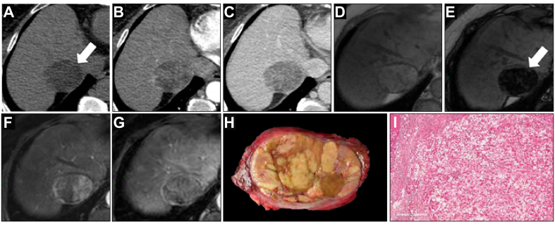
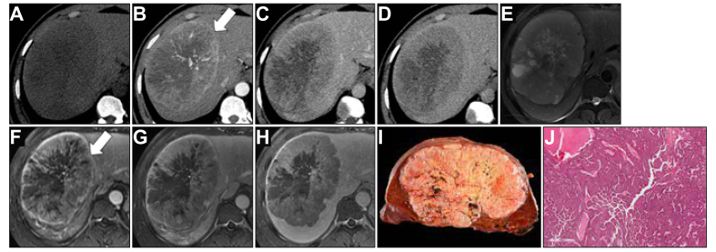
Two hundred and seventy-seven patients (median age 64.0 years, 215 [77.6%] men) were analysed, which involved 295 HCCs. There were 197 (66.7%) NOS-HCCs, 62 (21.0%) SH-HCCs, 23 (7.8%) MTM-HCCs, and 13 (4.5%) other rare subtypes. The following features were more frequent in MTM-HCC: elevated α-foetoprotein serum levels ( <0.001), tumour-in-vein ( <0.001 on CT, ≤0.052 on MRI), presence of at least 1 LR-M feature ( ≤0.010 on CT), infiltrative appearance ( ≤0.032 on CT), necrosis or severe ischaemia ( ≤0.038 on CT), and larger size ( ≤0.006 on CT, ≤0.011 on MRI). SH-HCC was associated with fat in mass ( <0.001 on CT, ≤0.002 on MRI). The distribution of the LI-RADS major features and categories in high-risk patients did not significantly differ among the 3 main HCC subtypes.
The distribution of LI-RADS major features and categories is not different among the HCC subtypes. Nevertheless, careful analysis of tumour-in-vein, LR-M, and ancillary features as well as clinico-biological data can provide information for the non-invasive diagnosis of HCC subtypes.
In high-risk patients, the overall distribution of LI-RADS major features and categories is not different among the histological subtypes of hepatocellular carcinoma, but tumour-in-vein, presence of LR-M features, and ancillary features can provide information for the non-invasive diagnosis of hepatocellular carcinoma subtypes.
肝细胞癌(HCC)的组织病理学亚型与不同的临床特征和预后相关。本研究旨在报告在一组切除肿瘤中,肝脏影像报告和数据系统(LI-RADS)定义的不同HCC亚型的影像特征,并评估HCC亚型对高危患者亚组中计算机断层扫描(CT)/磁共振成像(MRI)LI-RADS分类的影响。
这项经机构审查委员会批准的回顾性研究纳入了接受HCC切除且有可用组织病理学分类的患者。三名放射科医生独立回顾术前CT和MRI检查。阅片者根据LI-RADS v2018定义评估影像特征的存在情况,并为HCC高危患者提供LI-RADS分类。对未另作说明(NOS-HCC)、脂肪性肝炎相关性(SH-HCC)和大小梁-巨块型(MTM-HCC)HCC的LI-RADS特征和分类差异进行评估。
分析了277例患者(中位年龄64.0岁,215例[77.6%]为男性),共涉及295个HCC。其中有197个(66.7%)NOS-HCC、62个(21.0%)SH-HCC、23个(7.8%)MTM-HCC和13个(4.5%)其他罕见亚型。MTM-HCC中以下特征更为常见:血清甲胎蛋白水平升高(<0.001)、静脉内肿瘤(CT上<0.001,MRI上≤0.052)、至少存在1个LR-M特征(CT上≤0.010)、浸润性表现(CT上≤0.032)、坏死或严重缺血(CT上≤0.038)以及更大的尺寸(CT上≤0.006,MRI上≤0.011)。SH-HCC与肿块内脂肪有关(CT上<0.001,MRI上≤0.002)。高危患者中LI-RADS主要特征和分类的分布在3种主要HCC亚型之间无显著差异。
LI-RADS主要特征和分类的分布在HCC亚型之间无差异。然而,仔细分析静脉内肿瘤、LR-M和辅助特征以及临床生物学数据可为HCC亚型的无创诊断提供信息。
在高危患者中,肝细胞癌组织学亚型之间LI-RADS主要特征和分类的总体分布无差异,但静脉内肿瘤、LR-M特征的存在以及辅助特征可为肝细胞癌亚型的无创诊断提供信息。