• 文献检索
  • 文档翻译
  • 深度研究
  • 学术资讯
  • Suppr Zotero 插件Zotero 插件
  • 邀请有礼
  • 套餐&价格
  • 历史记录
应用&插件
Suppr Zotero 插件Zotero 插件浏览器插件Mac 客户端Windows 客户端微信小程序
定价
高级版会员购买积分包购买API积分包
服务
文献检索文档翻译深度研究API 文档MCP 服务
关于我们
关于 Suppr公司介绍联系我们用户协议隐私条款
关注我们

Suppr 超能文献

核心技术专利:CN118964589B侵权必究
粤ICP备2023148730 号-1Suppr @ 2026

文献检索

告别复杂PubMed语法,用中文像聊天一样搜索,搜遍4000万医学文献。AI智能推荐,让科研检索更轻松。

立即免费搜索

文件翻译

保留排版,准确专业,支持PDF/Word/PPT等文件格式,支持 12+语言互译。

免费翻译文档

深度研究

AI帮你快速写综述,25分钟生成高质量综述,智能提取关键信息,辅助科研写作。

立即免费体验

用于三维组织工程模型中微转移研究的鸡胚实验平台:癌症生物学、药物开发及纳米技术应用

Chick Embryo Experimental Platform for Micrometastases Research in a 3D Tissue Engineering Model: Cancer Biology, Drug Development, and Nanotechnology Applications.

作者信息

Guller Anna, Kuschnerus Inga, Rozova Vlada, Nadort Annemarie, Yao Yin, Khabir Zahra, Garcia-Bennett Alfonso, Liang Liuen Olivia, Polikarpova Aleksandra, Qian Yi, Goldys Ewa M, Zvyagin Andrei V

机构信息

Faculty of Science and Engineering, Macquarie University, Sydney, NSW 2109, Australia.

ARC Centre of Excellence for Nanoscale Biophotonics, Macquarie University, Sydney, NSW 2109, Australia.

出版信息

Biomedicines. 2021 Oct 29;9(11):1578. doi: 10.3390/biomedicines9111578.

DOI:10.3390/biomedicines9111578
PMID:34829808
原文链接:https://pmc.ncbi.nlm.nih.gov/articles/PMC8615510/
Abstract

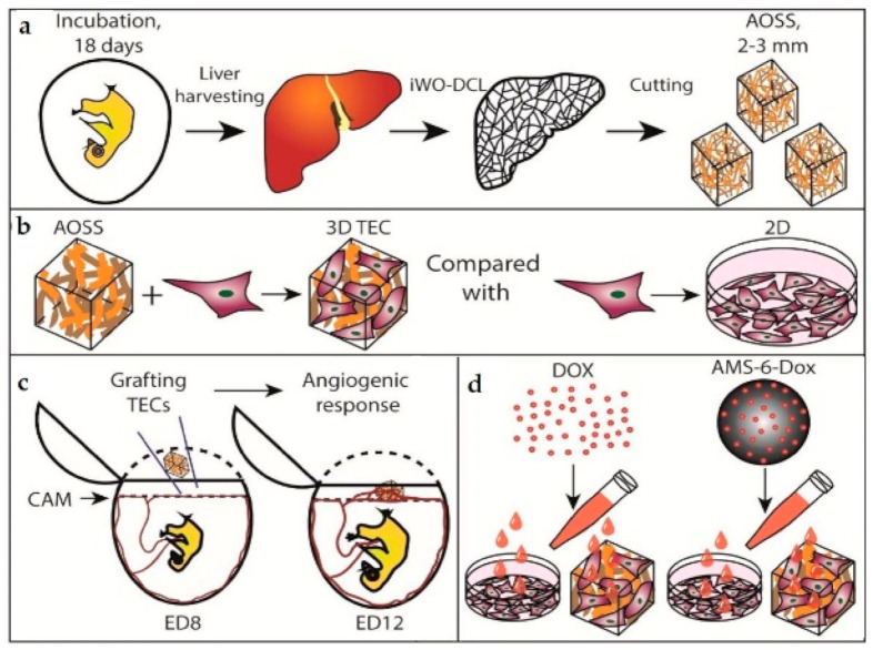
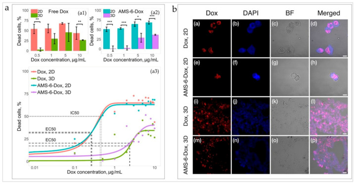
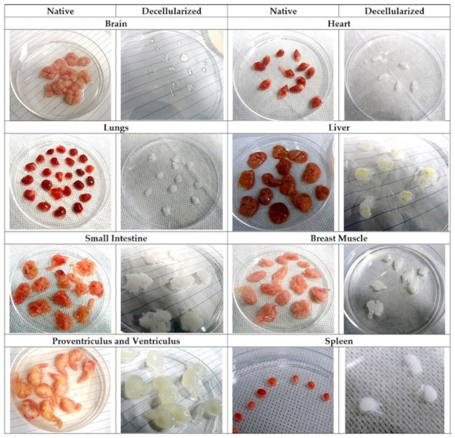
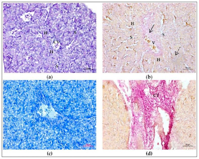
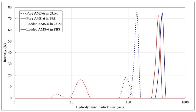
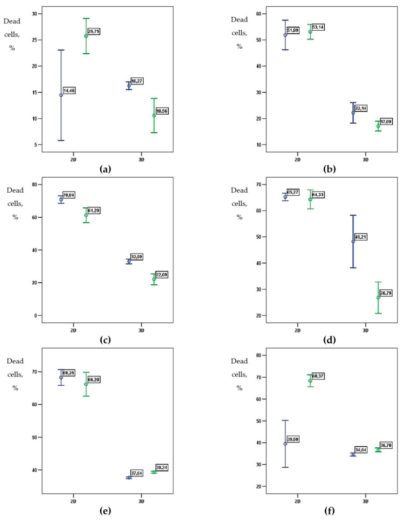
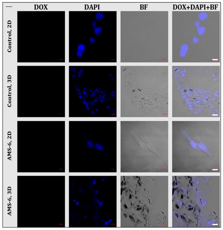
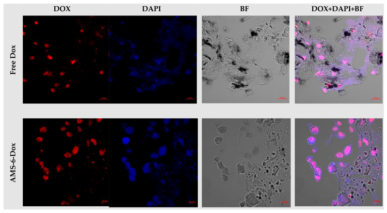
Colonization of distant organs by tumor cells is a critical step of cancer progression. The initial avascular stage of this process (micrometastasis) remains almost inaccessible to study due to the lack of relevant experimental approaches. Herein, we introduce an in vitro/in vivo model of organ-specific micrometastases of triple-negative breast cancer (TNBC) that is fully implemented in a cost-efficient chick embryo (CE) experimental platform. The model was built as three-dimensional (3D) tissue engineering constructs (TECs) combining human MDA-MB-231 cells and decellularized CE organ-specific scaffolds. TNBC cells colonized CE organ-specific scaffolds in 2-3 weeks, forming tissue-like structures. The feasibility of this methodology for basic cancer research, drug development, and nanomedicine was demonstrated on a model of hepatic micrometastasis of TNBC. We revealed that MDA-MB-231 differentially colonize parenchymal and stromal compartments of the liver-specific extracellular matrix (LS-ECM) and become more resistant to the treatment with molecular doxorubicin (Dox) and Dox-loaded mesoporous silica nanoparticles than in monolayer cultures. When grafted on CE chorioallantoic membrane, LS-ECM-based TECs induced angiogenic switch. These findings may have important implications for the diagnosis and treatment of TNBC. The methodology established here is scalable and adaptable for pharmacological testing and cancer biology research of various metastatic and primary tumors.

摘要

肿瘤细胞在远处器官的定植是癌症进展的关键步骤。由于缺乏相关实验方法,这一过程的初始无血管阶段(微转移)几乎无法进行研究。在此,我们介绍一种三阴性乳腺癌(TNBC)器官特异性微转移的体外/体内模型,该模型在经济高效的鸡胚(CE)实验平台上全面实现。该模型构建为三维(3D)组织工程构建体(TEC),将人MDA-MB-231细胞与脱细胞CE器官特异性支架相结合。TNBC细胞在2至3周内定植于CE器官特异性支架,形成组织样结构。在TNBC肝微转移模型上证明了该方法在基础癌症研究、药物开发和纳米医学方面的可行性。我们发现,MDA-MB-231在肝特异性细胞外基质(LS-ECM)的实质和间质区室中差异定植,并且与单层培养相比,对分子阿霉素(Dox)和载Dox的介孔二氧化硅纳米颗粒治疗更具抗性。当移植到CE尿囊膜上时,基于LS-ECM的TEC诱导血管生成转换。这些发现可能对TNBC的诊断和治疗具有重要意义。这里建立的方法可扩展并适用于各种转移性和原发性肿瘤的药理测试和癌症生物学研究。

https://cdn.ncbi.nlm.nih.gov/pmc/blobs/cd6f/8615510/3d4e765e3429/biomedicines-09-01578-g008.jpg
https://cdn.ncbi.nlm.nih.gov/pmc/blobs/cd6f/8615510/d8feb4ddc4b3/biomedicines-09-01578-g0A1.jpg
https://cdn.ncbi.nlm.nih.gov/pmc/blobs/cd6f/8615510/fed9a289060e/biomedicines-09-01578-g0A2.jpg
https://cdn.ncbi.nlm.nih.gov/pmc/blobs/cd6f/8615510/657e8ca6527f/biomedicines-09-01578-g0A3.jpg
https://cdn.ncbi.nlm.nih.gov/pmc/blobs/cd6f/8615510/ed3677bcaeac/biomedicines-09-01578-g0A4.jpg
https://cdn.ncbi.nlm.nih.gov/pmc/blobs/cd6f/8615510/8de240df1a9a/biomedicines-09-01578-g0A5.jpg
https://cdn.ncbi.nlm.nih.gov/pmc/blobs/cd6f/8615510/943d7cdeb437/biomedicines-09-01578-g0A6.jpg
https://cdn.ncbi.nlm.nih.gov/pmc/blobs/cd6f/8615510/e2b65247baac/biomedicines-09-01578-g0A7.jpg
https://cdn.ncbi.nlm.nih.gov/pmc/blobs/cd6f/8615510/96f747f2c484/biomedicines-09-01578-g0A8.jpg
https://cdn.ncbi.nlm.nih.gov/pmc/blobs/cd6f/8615510/c01e126da037/biomedicines-09-01578-g0A9.jpg
https://cdn.ncbi.nlm.nih.gov/pmc/blobs/cd6f/8615510/8071f8cea4aa/biomedicines-09-01578-g0A10.jpg
https://cdn.ncbi.nlm.nih.gov/pmc/blobs/cd6f/8615510/c9324531fe6e/biomedicines-09-01578-g0A11.jpg
https://cdn.ncbi.nlm.nih.gov/pmc/blobs/cd6f/8615510/0dc90f876d0c/biomedicines-09-01578-g0A12.jpg
https://cdn.ncbi.nlm.nih.gov/pmc/blobs/cd6f/8615510/acdfc89e2d9f/biomedicines-09-01578-g0A13.jpg
https://cdn.ncbi.nlm.nih.gov/pmc/blobs/cd6f/8615510/8daa27b34996/biomedicines-09-01578-g0A14.jpg
https://cdn.ncbi.nlm.nih.gov/pmc/blobs/cd6f/8615510/ba8a126e6d64/biomedicines-09-01578-g0A15.jpg
https://cdn.ncbi.nlm.nih.gov/pmc/blobs/cd6f/8615510/6d6a4d4b0138/biomedicines-09-01578-g0A16.jpg
https://cdn.ncbi.nlm.nih.gov/pmc/blobs/cd6f/8615510/9b949eecb1e3/biomedicines-09-01578-g0A17.jpg
https://cdn.ncbi.nlm.nih.gov/pmc/blobs/cd6f/8615510/498db4ef4231/biomedicines-09-01578-g0A18.jpg
https://cdn.ncbi.nlm.nih.gov/pmc/blobs/cd6f/8615510/24b7d9dc817f/biomedicines-09-01578-g0A19.jpg
https://cdn.ncbi.nlm.nih.gov/pmc/blobs/cd6f/8615510/2928f297f403/biomedicines-09-01578-g0A20.jpg
https://cdn.ncbi.nlm.nih.gov/pmc/blobs/cd6f/8615510/e627839e0b59/biomedicines-09-01578-g0A21.jpg
https://cdn.ncbi.nlm.nih.gov/pmc/blobs/cd6f/8615510/5c8cb71ba2b7/biomedicines-09-01578-g0A22.jpg
https://cdn.ncbi.nlm.nih.gov/pmc/blobs/cd6f/8615510/a627079a7860/biomedicines-09-01578-g0A23.jpg
https://cdn.ncbi.nlm.nih.gov/pmc/blobs/cd6f/8615510/89d8263763ef/biomedicines-09-01578-g0A24.jpg
https://cdn.ncbi.nlm.nih.gov/pmc/blobs/cd6f/8615510/9fdbbe26b6af/biomedicines-09-01578-g001.jpg
https://cdn.ncbi.nlm.nih.gov/pmc/blobs/cd6f/8615510/d6475add5201/biomedicines-09-01578-g002.jpg
https://cdn.ncbi.nlm.nih.gov/pmc/blobs/cd6f/8615510/ef706c69399c/biomedicines-09-01578-g003.jpg
https://cdn.ncbi.nlm.nih.gov/pmc/blobs/cd6f/8615510/8af94c6958fd/biomedicines-09-01578-g004.jpg
https://cdn.ncbi.nlm.nih.gov/pmc/blobs/cd6f/8615510/f9acb7ecdaef/biomedicines-09-01578-g005.jpg
https://cdn.ncbi.nlm.nih.gov/pmc/blobs/cd6f/8615510/fd0885ef04db/biomedicines-09-01578-g006.jpg
https://cdn.ncbi.nlm.nih.gov/pmc/blobs/cd6f/8615510/57af206ca66c/biomedicines-09-01578-g007.jpg
https://cdn.ncbi.nlm.nih.gov/pmc/blobs/cd6f/8615510/3d4e765e3429/biomedicines-09-01578-g008.jpg

相似文献

1
Chick Embryo Experimental Platform for Micrometastases Research in a 3D Tissue Engineering Model: Cancer Biology, Drug Development, and Nanotechnology Applications.用于三维组织工程模型中微转移研究的鸡胚实验平台:癌症生物学、药物开发及纳米技术应用
Biomedicines. 2021 Oct 29;9(11):1578. doi: 10.3390/biomedicines9111578.
2
Breast Cancer Cell Type and Biomechanical Properties of Decellularized Mouse Organs Drives Tumor Cell Colonization.乳腺癌细胞类型和去细胞化小鼠器官的生物力学特性驱动肿瘤细胞定植。
Cells. 2023 Aug 9;12(16):2030. doi: 10.3390/cells12162030.
3
Macrophage-Derived Extracellular Vesicles as Drug Delivery Systems for Triple Negative Breast Cancer (TNBC) Therapy.巨噬细胞衍生的细胞外囊泡作为三阴性乳腺癌 (TNBC) 治疗的药物递送系统。
J Neuroimmune Pharmacol. 2020 Sep;15(3):487-500. doi: 10.1007/s11481-019-09884-9. Epub 2019 Nov 13.
4
Synthesis of micellar-like terpolymer nanoparticles with reductively-cleavable cross-links and evaluation of efficacy in 2D and 3D models of triple negative breast cancer.具有还原可裂解交联的胶束状三元共聚物纳米粒子的合成及其在三阴性乳腺癌 2D 和 3D 模型中的功效评价。
J Control Release. 2020 Jul 10;323:549-564. doi: 10.1016/j.jconrel.2020.04.049. Epub 2020 May 3.
5
Sequentially-targeted biomimetic nano drug system for triple-negative breast cancer ablation and lung metastasis inhibition.用于三阴性乳腺癌消融和肺转移抑制的顺序靶向仿生纳米药物系统
Acta Biomater. 2020 Sep 1;113:554-569. doi: 10.1016/j.actbio.2020.06.025. Epub 2020 Jun 20.
6
Dual-targeted hybrid nanoparticles of synergistic drugs for treating lung metastases of triple negative breast cancer in mice.用于治疗小鼠三阴性乳腺癌肺转移的协同药物双靶点混合纳米颗粒。
Acta Pharmacol Sin. 2017 Jun;38(6):835-847. doi: 10.1038/aps.2016.166. Epub 2017 Feb 20.
7
Hyaluronic acid-capped compact silica-supported mesoporous titania nanoparticles for ligand-directed delivery of doxorubicin.透明质酸封端的紧密型硅基介孔二氧化钛纳米粒子用于阿霉素的配体导向递送。
Acta Biomater. 2018 Oct 15;80:364-377. doi: 10.1016/j.actbio.2018.09.006. Epub 2018 Sep 8.
8
Combined mTOR inhibitor rapamycin and doxorubicin-loaded cyclic octapeptide modified liposomes for targeting integrin α3 in triple-negative breast cancer.载多柔比星和 mTOR 抑制剂雷帕霉素的环八肽修饰脂质体靶向三阴性乳腺癌整合素 α3。
Biomaterials. 2014 Jul;35(20):5347-5358. doi: 10.1016/j.biomaterials.2014.03.036. Epub 2014 Apr 13.
9
Development of an ex vivo breast cancer lung colonization model utilizing a decellularized lung matrix.利用去细胞肺基质建立体外乳腺癌肺转移模型。
Integr Biol (Camb). 2015 Dec;7(12):1518-25. doi: 10.1039/c5ib00157a. Epub 2015 Nov 13.
10
Delivery of thymoquinone through hyaluronic acid-decorated mixed Pluronic® nanoparticles to attenuate angiogenesis and metastasis of triple-negative breast cancer.通过透明质酸修饰的混合 Pluronic®纳米粒子传递百里醌来抑制三阴性乳腺癌的血管生成和转移。
J Control Release. 2020 Jun 10;322:357-374. doi: 10.1016/j.jconrel.2020.03.033. Epub 2020 Mar 31.

引用本文的文献

1
Breast cancer metastasis progression is associated with elevated activity of kynurenine monooxygenase and kynureninase.乳腺癌转移进展与犬尿氨酸单加氧酶和犬尿氨酸酶活性升高有关。
Br J Cancer. 2024 Dec;131(12):1881-1892. doi: 10.1038/s41416-024-02889-z. Epub 2024 Nov 18.
2
The Chorioallantoic Membrane Xenograft Assay as a Reliable Model for Investigating the Biology of Breast Cancer.绒毛膜尿囊膜异种移植试验作为研究乳腺癌生物学特性的可靠模型
Cancers (Basel). 2023 Mar 10;15(6):1704. doi: 10.3390/cancers15061704.
3
Impact of intestinal dysbiosis on breast cancer metastasis and progression.

本文引用的文献

1
Machine learning reveals mesenchymal breast carcinoma cell adaptation in response to matrix stiffness.机器学习揭示了间质型乳腺癌细胞对基质硬度的适应。
PLoS Comput Biol. 2021 Jul 23;17(7):e1009193. doi: 10.1371/journal.pcbi.1009193. eCollection 2021 Jul.
2
Liver metastases.肝转移。
Nat Rev Dis Primers. 2021 Apr 15;7(1):27. doi: 10.1038/s41572-021-00261-6.
3
Oxygen-Carrying Polymer Nanoconstructs for Radiodynamic Therapy of Deep Hypoxic Malignant Tumors.用于深部缺氧恶性肿瘤放射动力治疗的载氧聚合物纳米结构
肠道菌群失调对乳腺癌转移和进展的影响。
Front Oncol. 2022 Nov 7;12:1037831. doi: 10.3389/fonc.2022.1037831. eCollection 2022.
Biomedicines. 2021 Mar 22;9(3):322. doi: 10.3390/biomedicines9030322.
4
The matrix in cancer.癌症中的基质。
Nat Rev Cancer. 2021 Apr;21(4):217-238. doi: 10.1038/s41568-020-00329-7. Epub 2021 Feb 15.
5
The lingering mysteries of metastatic recurrence in breast cancer.乳腺癌转移复发的未解之谜。
Br J Cancer. 2021 Jan;124(1):13-26. doi: 10.1038/s41416-020-01161-4. Epub 2020 Nov 26.
6
A Complex and Evolutive Character: Two Face Aspects of ECM in Tumor Progression.一个复杂且不断演变的特征:肿瘤进展中细胞外基质的两个方面
Front Oncol. 2020 Aug 28;10:1620. doi: 10.3389/fonc.2020.01620. eCollection 2020.
7
Exosomal MicroRNAs and Organotropism in Breast Cancer Metastasis.外泌体微小RNA与乳腺癌转移中的器官嗜性
Cancers (Basel). 2020 Jul 7;12(7):1827. doi: 10.3390/cancers12071827.
8
Acellular matrix hydrogel for repair of the temporomandibular joint disc.细胞外基质水凝胶修复颞下颌关节盘。
J Biomed Mater Res B Appl Biomater. 2020 Oct;108(7):2995-3007. doi: 10.1002/jbm.b.34629. Epub 2020 Jun 29.
9
Decellularized Extracellular Matrix for Bioengineering Physiomimetic 3D in Vitro Tumor Models.去细胞细胞外基质用于生物工程仿生 3D 体外肿瘤模型。
Trends Biotechnol. 2020 Dec;38(12):1397-1414. doi: 10.1016/j.tibtech.2020.04.006. Epub 2020 May 13.
10
Framing cancer progression: influence of the organ- and tumour-specific matrisome.构建癌症进展模型:器官和肿瘤特异性基质的影响。
FEBS J. 2020 Apr;287(8):1454-1477. doi: 10.1111/febs.15223. Epub 2020 Feb 6.