Laboratory of Molecular Neurobiology, Academy of Biology and Biotechnology, Southern Federal University, pr. Stachki 194/1, 344090 Rostov-on-Don, Russia.
Int J Mol Sci. 2021 Nov 19;22(22):12483. doi: 10.3390/ijms222212483.
Cerebral ischemia, a common cerebrovascular disease, is one of the great threats to human health and new targets for stroke therapy are needed. The transcriptional activity in the cell is regulated by epigenetic processes such as DNA methylation/demethylation, acetylation/deacetylation, histone methylation, etc. Changes in DNA methylation after ischemia can have both neuroprotective and neurotoxic effects depending on the degree of ischemia damage, the time elapsed after injury, and the site of methylation.
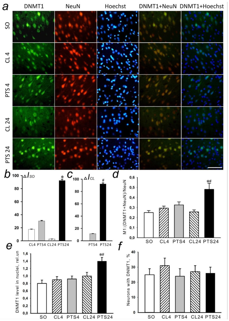
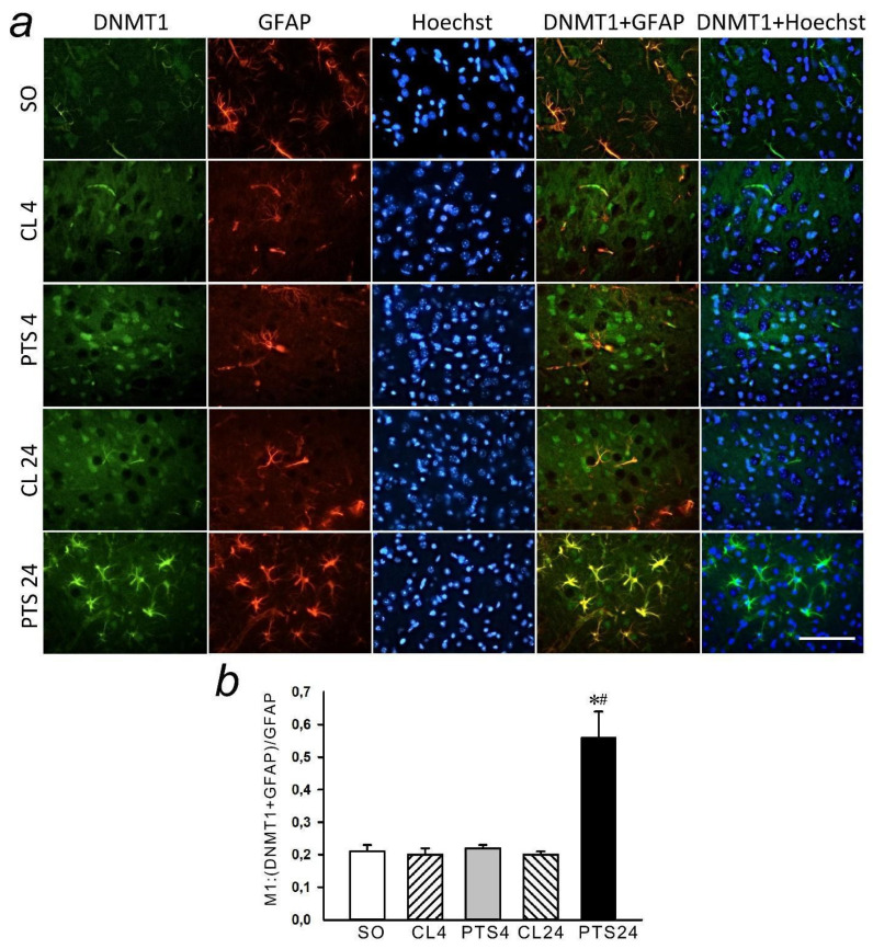
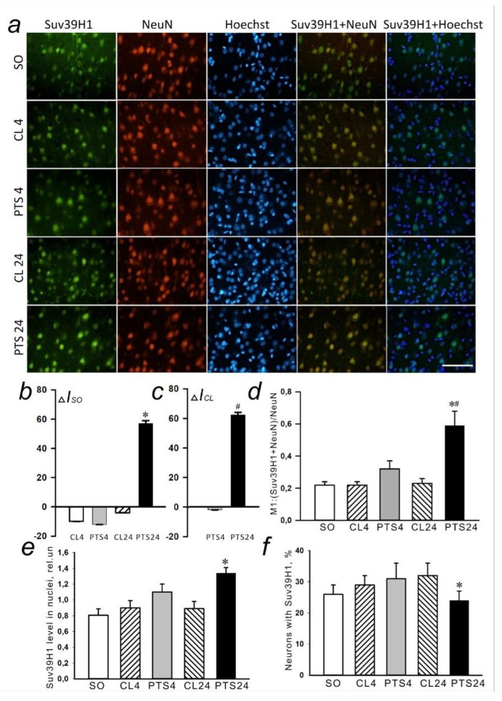
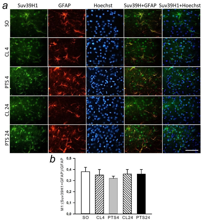
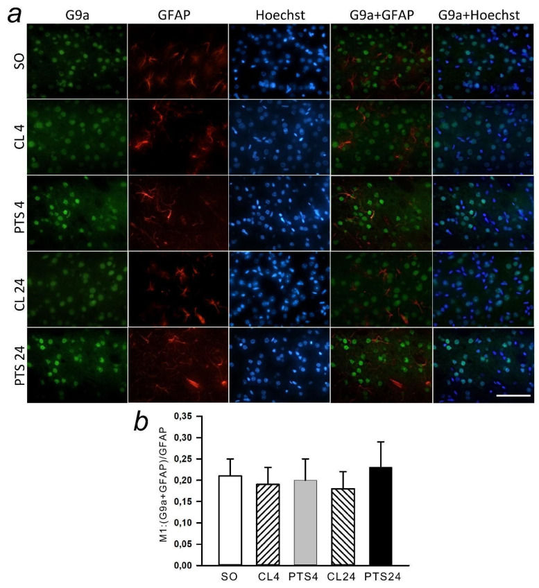
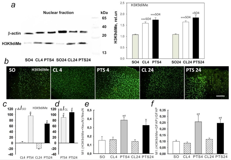
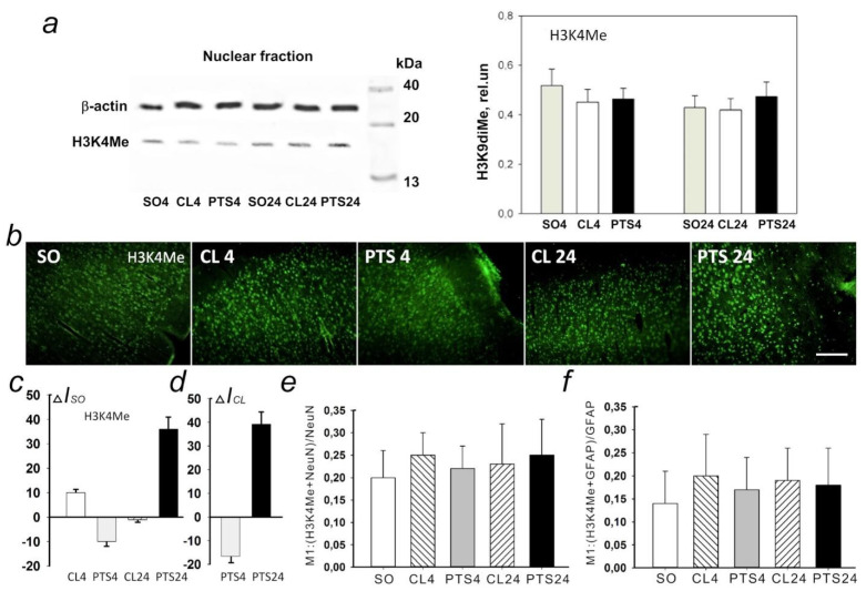
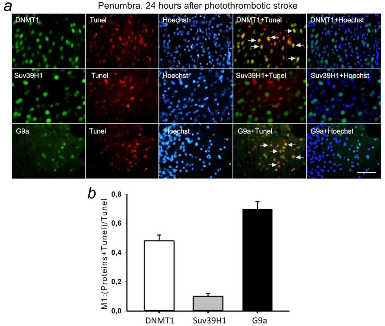
In this study, we investigated the changes in the expression and intracellular localization of DNA methyltransferase DNMT1, histone methyltransferases SUV39H1, and G9a in penumbra neurons and astrocytes at 4 and 24 h after stroke in the rat cerebral cortex using photothrombotic stroke (PTS) model. Methods of immunofluorescence microscopy analysis, apoptosis analysis, and immunoblotting were used. Additionally, we have studied the effect of DNMT1 and G9a inhibitors on the volume of PTS-induced infarction and apoptosis of penumbra cells in the cortex of mice after PTS.
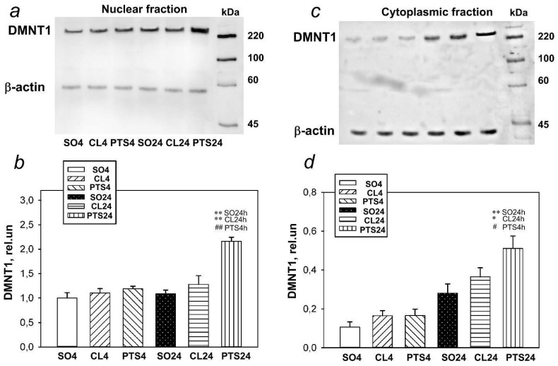
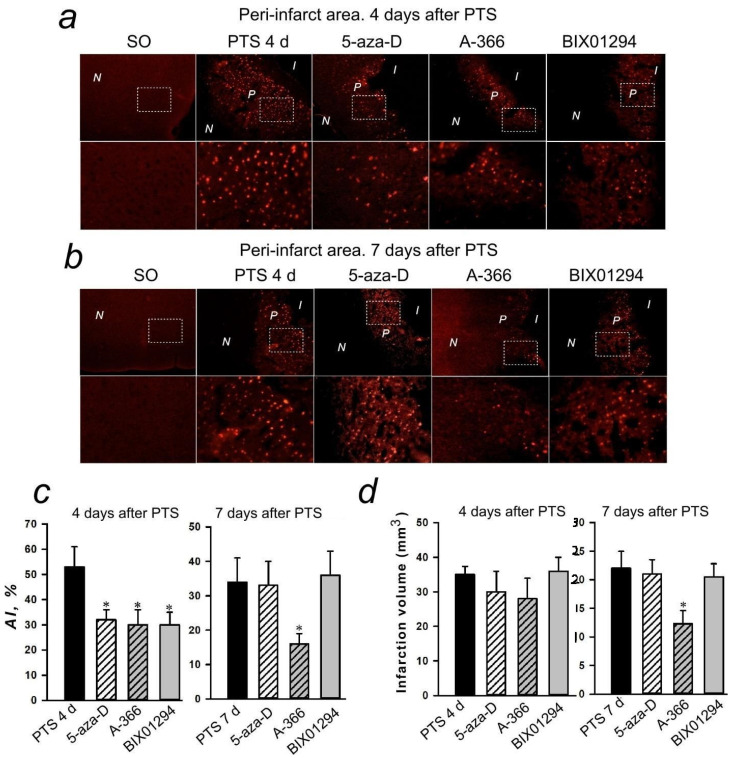
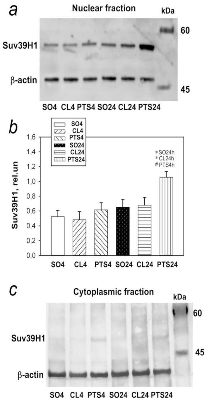
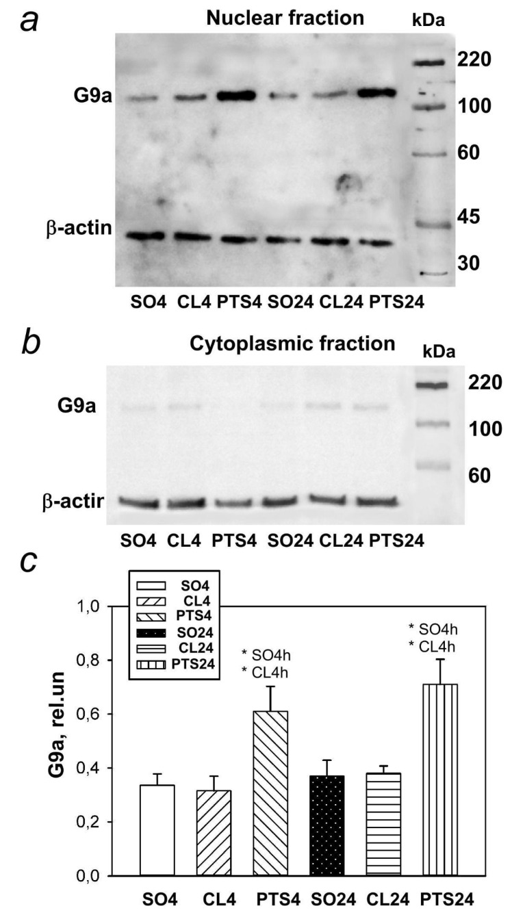
This study has shown that the level of DNMT1 increased in the nuclear and cytoplasmic fractions of the penumbra tissue at 24 h after PTS. Inhibition of DNMT1 by 5-aza-2'-deoxycytidine protected cells of PTS-induced penumbra from apoptosis. An increase in the level of SUV39H1 in the penumbra was found at 24 h after PTS and G9a was overexpressed at 4 and 24 h after PTS. G9a inhibitors A-366 and BIX01294 protected penumbra cells from apoptosis and reduced the volume of PTS-induced cerebral infarction.
Thus, the data obtained show that DNA methyltransferase DNMT1 and histone methyltransferase G9a can be potential protein targets in ischemic penumbra cells, and their inhibitors are potential neuroprotective agents capable of protecting penumbra cells from postischemic damage to the cerebral cortex.
脑缺血是一种常见的脑血管疾病,是人类健康的重大威胁之一,因此需要寻找新的脑卒中治疗靶点。细胞中的转录活性受表观遗传过程的调节,如 DNA 甲基化/去甲基化、乙酰化/去乙酰化、组蛋白甲基化等。缺血后 DNA 甲基化的变化可产生神经保护和神经毒性作用,这取决于缺血损伤的程度、损伤后时间的长短以及甲基化的部位。
在这项研究中,我们使用光血栓性脑卒中(PTS)模型研究了大鼠大脑皮质脑卒中后 4 小时和 24 小时缺血半影区神经元和星形胶质细胞中 DNA 甲基转移酶 DNMT1、组蛋白甲基转移酶 SUV39H1 和 G9a 的表达和细胞内定位的变化。使用免疫荧光显微镜分析、凋亡分析和免疫印迹法。此外,我们还研究了 DNMT1 和 G9a 抑制剂对 PTS 后小鼠大脑皮质 PTS 诱导的梗死体积和缺血半影区细胞凋亡的影响。
本研究表明,PTS 后 24 小时,DNMT1 水平在缺血半影区组织的核和胞质部分增加。用 5-氮杂-2'-脱氧胞苷抑制 DNMT1 可使 PTS 诱导的缺血半影区细胞免于凋亡。PTS 后 24 小时发现 SUV39H1 水平增加,PTS 后 4 小时和 24 小时 G9a 过表达。G9a 抑制剂 A-366 和 BIX01294 可保护缺血半影区细胞免于凋亡,并减少 PTS 诱导的脑梗死体积。
因此,研究结果表明,DNA 甲基转移酶 DNMT1 和组蛋白甲基转移酶 G9a 可能是缺血半影区细胞的潜在蛋白靶点,其抑制剂可能是保护缺血半影区细胞免受大脑皮质缺血后损伤的潜在神经保护剂。