Bauer Thomas, Gubi Daniela, Klufa Jörg, Novoszel Philipp, Holcmann Martin, Sibilia Maria
Department of Medicine I, Institute of Cancer Research, Medical University of Vienna and Comprehensive Cancer Center, 1090 Vienna, Austria.
Life (Basel). 2021 Nov 16;11(11):1237. doi: 10.3390/life11111237.
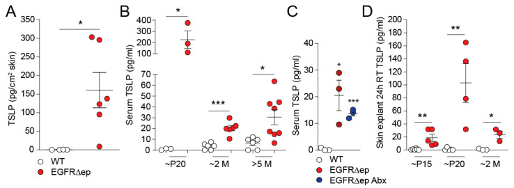
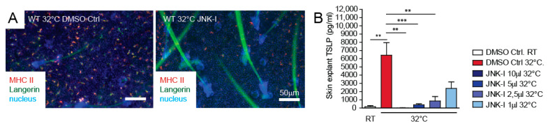
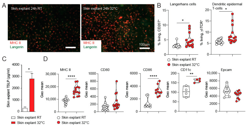
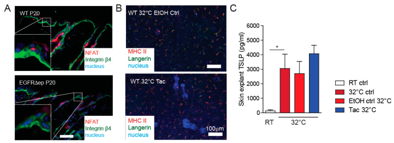
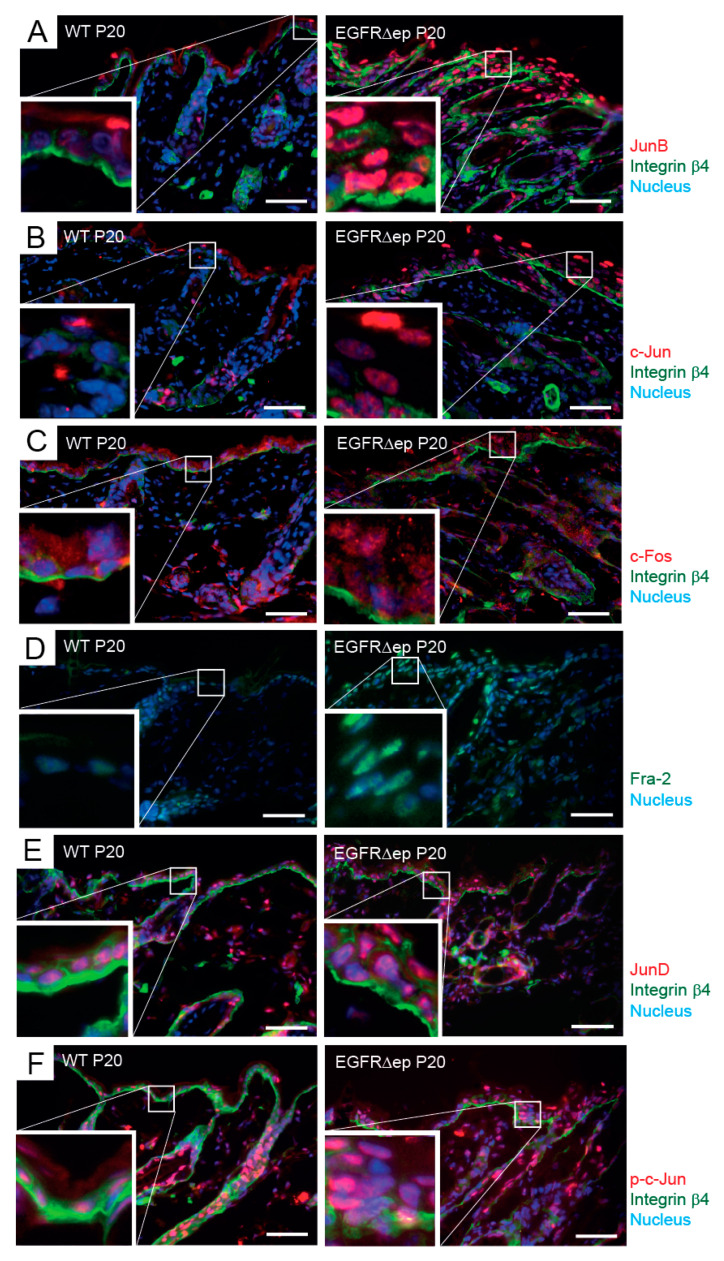
The skin is the outermost barrier protecting the body from pathogenic invasion and environmental insults. Its breakdown initiates the start of skin inflammation. The epidermal growth factor (EGFR) on keratinocytes protects this barrier, and its dysfunction leads to atopic dermatitis-like skin disease. One of the initial cytokines expressed upon skin barrier breach and during atopic dermatitis is TSLP. Here, we describe the expression and secretion of TSLP during EGFR inhibition and present an ex-vivo model, which mimics the early events after barrier insult. Skin explants floated on culture medium at 32 °C released TSLP in parallel to the activation of the resident Langerhans cell network. We could further show the up-regulation and activation of the AP-1 family of transcription factors during atopic-like skin inflammation and its involvement in TSLP production from the skin explant cultures. Inhibition of the c-Jun N-terminal kinase pathway led to a dose-dependent blunting of TSLP release. These data indicate the involvement of AP-1 during the early stages of atopic-like skin inflammation and highlight a novel therapeutic approach by targeting it. Therefore, skin explant cultures mimic the early events during skin barrier immunity and provide a suitable model to test therapeutic intervention.
皮肤是保护身体免受病原体入侵和环境侵害的最外层屏障。其破损会引发皮肤炎症。角质形成细胞上的表皮生长因子(EGFR)可保护这一屏障,其功能障碍会导致特应性皮炎样皮肤病。在皮肤屏障破坏时以及特应性皮炎期间最初表达的细胞因子之一是TSLP。在此,我们描述了EGFR抑制过程中TSLP的表达和分泌,并提出了一种体外模型,该模型模拟屏障受损后的早期事件。漂浮在32℃培养基上的皮肤外植体释放TSLP的情况与驻留朗格汉斯细胞网络的激活情况平行。我们还能进一步证明在特应样皮肤炎症期间转录因子AP-1家族的上调和激活及其参与皮肤外植体培养物中TSLP的产生。抑制c-Jun N端激酶途径会导致TSLP释放呈剂量依赖性减弱。这些数据表明AP-1参与了特应样皮肤炎症的早期阶段,并突出了一种通过靶向它的新型治疗方法。因此,皮肤外植体培养物模拟了皮肤屏障免疫期间的早期事件,并提供了一个测试治疗干预的合适模型。