Shu Bo, Zhou Ying-Xia, Li Hao, Zhang Rui-Zhi, He Chao, Yang Xin
Department of General Surgery, The Second Xiangya Hospital, Central South University, 410011, Changsha, Hunan Province, China.
Department of Surgical Operation, The Second Xiangya Hospital, Central South University, 410011, Changsha, Hunan Province, China.
Cell Death Discov. 2021 Nov 27;7(1):368. doi: 10.1038/s41420-021-00756-x.
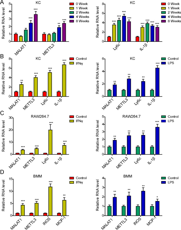
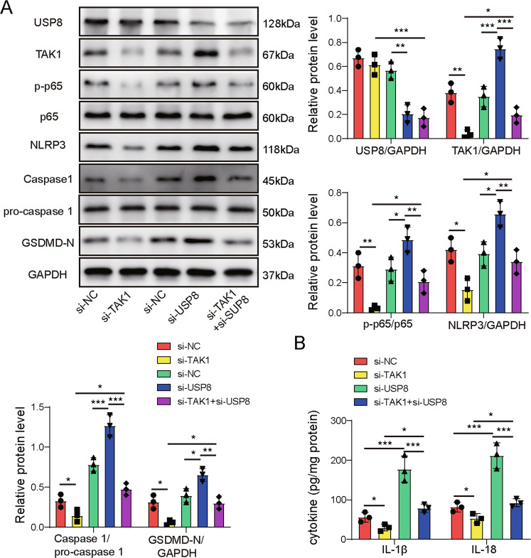
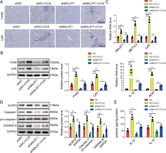
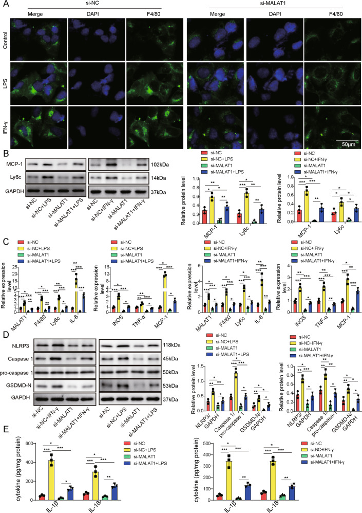
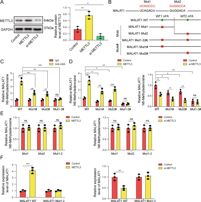
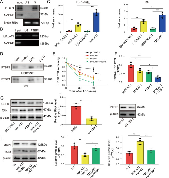
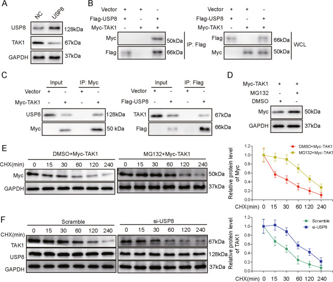
Pro-inflammatory M1 macrophages, via activating hepatic stellate cells, contribute to liver fibrosis. In this study, we examined the mechanism and the significance of a signaling axis, METTL3/MALAT1/PTBP1/USP8/TAK1, in regulating pyroptosis and M1 polarization of hepatic macrophages. Liver fibrosis model was established in vivo by CCl treatment; M1 polarization was induced in vitro by treating macrophages with lipopolysaccharide or interferon γ. Expressions of METTL3, MALAT1, PTBP1, USP8, and TAK1 were measured by RT-PCR and/or Western blot in Kupffer cells (KCs) isolated from in vivo model or in vitro activated macrophages. Macrophage phenotypes including inflammation (RT-qPCR analysis of a panel of proinflammatory cytokines and ELISA on productions of interleukin (IL)-1β and IL-18) and pyroptosis (Western blot of NLRP3, Caspase-1, and GSDMD) were investigated. The impact of METTL3 on mA methylation of MALAT1 was examined by methylated RNA immunoprecipitation (RIP), the interaction between PTBP1 and MALAT1 or USP8 mRNA by combining RNA pull-down, RIP, and RNA stability assays, and the crosstalk between USP8 and TAK1 by co-immunoprecipitation and protein degradation assays. Functional significance of individual component of METTL3/MALAT1/PTBP1/USP8/TAK1 axis was assessed by combining gain-of-function and loss-of-function approaches. In KCs isolated from in vivo liver fibrosis model or in vitro M1-polarized macrophages, METTL3 was up-regulated, and sequentially, it increased MALAT1 level via mA methylation, which promoted USP8 mRNA degradation through the interaction with PTBP1. Reduced USP8 expression regulated the ubiquitination and protein stability of TAK1, which promoted pyroptosis and inflammation of macrophages. The signaling cascade METTL3/MALAT1/PTBP1/USP8/TAK1, by essentially stimulating pyroptosis and inflammation of macrophages, aggravates liver fibrosis. Therefore, targeting individual components of this axis may benefit the treatment of liver fibrosis.
促炎M1巨噬细胞通过激活肝星状细胞,促进肝纤维化。在本研究中,我们研究了信号轴METTL3/MALAT1/PTBP1/USP8/TAK1在调节肝巨噬细胞焦亡和M1极化中的机制及意义。通过CCl处理在体内建立肝纤维化模型;通过用脂多糖或干扰素γ处理巨噬细胞在体外诱导M1极化。通过RT-PCR和/或蛋白质印迹法检测从体内模型分离的库普弗细胞(KC)或体外活化巨噬细胞中METTL3、MALAT1、PTBP1、USP8和TAK1的表达。研究巨噬细胞表型,包括炎症(一组促炎细胞因子的RT-qPCR分析以及白细胞介素(IL)-1β和IL-18产生的ELISA)和焦亡(NLRP3、半胱天冬酶-1和GSDMD的蛋白质印迹)。通过甲基化RNA免疫沉淀(RIP)检测METTL3对MALAT1的m⁶A甲基化的影响,通过结合RNA下拉、RIP和RNA稳定性测定检测PTBP1与MALAT1或USP8 mRNA之间的相互作用,通过共免疫沉淀和蛋白质降解测定检测USP8与TAK1之间的串扰。通过结合功能获得和功能丧失方法评估METTL3/MALAT1/PTBP1/USP8/TAK1轴单个组分的功能意义。在从体内肝纤维化模型分离的KC或体外M1极化巨噬细胞中,METTL3上调,随后,它通过m⁶A甲基化增加MALAT1水平,这通过与PTBP1的相互作用促进USP8 mRNA降解。USP8表达降低调节TAK1的泛素化和蛋白质稳定性,从而促进巨噬细胞的焦亡和炎症。信号级联METTL3/MALAT1/PTBP1/USP8/TAK1通过本质上刺激巨噬细胞的焦亡和炎症,加重肝纤维化。因此,靶向该轴的单个组分可能有益于肝纤维化的治疗。