• 文献检索
  • 文档翻译
  • 深度研究
  • 学术资讯
  • Suppr Zotero 插件Zotero 插件
  • 邀请有礼
  • 套餐&价格
  • 历史记录
应用&插件
Suppr Zotero 插件Zotero 插件浏览器插件Mac 客户端Windows 客户端微信小程序
定价
高级版会员购买积分包购买API积分包
服务
文献检索文档翻译深度研究API 文档MCP 服务
关于我们
关于 Suppr公司介绍联系我们用户协议隐私条款
关注我们

Suppr 超能文献

核心技术专利:CN118964589B侵权必究
粤ICP备2023148730 号-1Suppr @ 2026

文献检索

告别复杂PubMed语法,用中文像聊天一样搜索,搜遍4000万医学文献。AI智能推荐,让科研检索更轻松。

立即免费搜索

文件翻译

保留排版,准确专业,支持PDF/Word/PPT等文件格式,支持 12+语言互译。

免费翻译文档

深度研究

AI帮你快速写综述,25分钟生成高质量综述,智能提取关键信息,辅助科研写作。

立即免费体验

新冠病毒检测呈阴性的幸存者中较高的边缘系统和基底神经节体积及其与疲劳的关系。

Higher Limbic and Basal Ganglia volumes in surviving COVID-negative patients and the relations to fatigue.

作者信息

Hafiz Rakibul, Gandhi Tapan Kumar, Mishra Sapna, Prasad Alok, Mahajan Vidur, Di Xin, Natelson Benjamin H, Biswal Bharat B

机构信息

Department of Biomedical Engineering, New Jersey Institute of Technology (NJIT), 323 Dr Martin Luther King Jr Blvd, Newark, NJ 07102, USA.

Department of Electrical Engineering, Indian Institute of Technology (IIT), Block II, IIT Delhi Main Rd, IIT Campus, Hauz Khas, New Delhi, Delhi 110016, India.

出版信息

medRxiv. 2022 Mar 1:2021.11.23.21266761. doi: 10.1101/2021.11.23.21266761.

DOI:10.1101/2021.11.23.21266761
PMID:34845462
原文链接:https://pmc.ncbi.nlm.nih.gov/articles/PMC8629206/
Abstract

BACKGROUND

Among systemic abnormalities caused by the novel coronavirus, little is known about the critical attack on the central nervous system (CNS). Few studies have shown cerebrovascular pathologies that indicate CNS involvement in acute patients. However, replication studies are necessary to verify if these effects persist in COVID-19 survivors more conclusively. Furthermore, recent studies indicate fatigue is highly prevalent among 'long-COVID' patients. How morphometry in each group relate to work-related fatigue need to be investigated.

METHOD

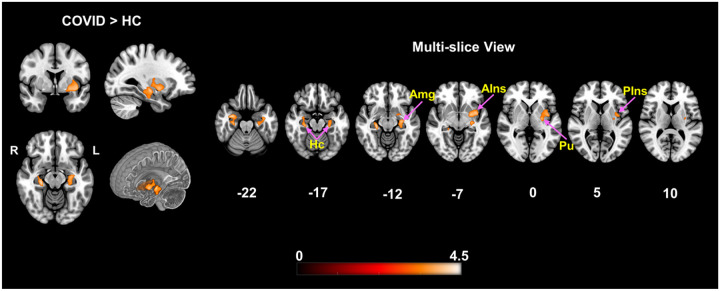
COVID survivors were MRI scanned two weeks after hospital discharge. We hypothesized, these survivors will demonstrate altered gray matter volume (GMV) and experience higher fatigue levels when compared to healthy controls, leading to stronger correlation of GMV with fatigue. Voxel-based morphometry was performed on T1-weighted MRI images between 46 survivors and 30 controls. Unpaired two-sample t-test and multiple linear regression were performed to observe group differences and correlation of fatigue with GMV.

RESULTS

The COVID group experienced significantly higher fatigue levels and GMV of this group was significantly higher within the and when compared to healthy controls. Moreover, while a significant positive correlation was observed across the whole group between GMV and self-reported fatigue, COVID subjects showed stronger effects within the and .

CONCLUSION

Brain regions with GMV alterations in our analysis align with both single case acute patient reports and current group level neuroimaging findings. We also newly report a stronger positive correlation of GMV with fatigue among COVID survivors within brain regions associated with fatigue, indicating a link between structural abnormality and brain function in this cohort.

摘要

背景

在新型冠状病毒引起的全身异常中,对中枢神经系统(CNS)的严重攻击了解甚少。很少有研究显示脑血管病变表明急性患者存在中枢神经系统受累。然而,需要进行重复研究以更确凿地验证这些影响在新冠病毒疾病(COVID-19)幸存者中是否持续存在。此外,最近的研究表明疲劳在“长期新冠”患者中非常普遍。需要研究每组的形态测量与工作相关疲劳之间的关系。

方法

新冠病毒疾病幸存者在出院两周后接受磁共振成像(MRI)扫描。我们假设,与健康对照组相比,这些幸存者将表现出灰质体积(GMV)改变,并经历更高水平的疲劳,从而导致GMV与疲劳之间的更强相关性。对46名幸存者和30名对照组的T1加权MRI图像进行基于体素的形态测量。进行非配对双样本t检验和多元线性回归以观察组间差异以及疲劳与GMV的相关性。

结果

新冠病毒疾病组经历了显著更高水平的疲劳,并且与健康对照组相比,该组的GMV在[具体脑区1]和[具体脑区2]内显著更高。此外,虽然在整个组中观察到GMV与自我报告的疲劳之间存在显著正相关,但新冠病毒疾病受试者在[具体脑区1]和[具体脑区2]内表现出更强的效应。

结论

我们分析中GMV改变的脑区与单例急性患者报告以及当前组水平的神经影像学发现一致。我们还首次报告了在与疲劳相关的脑区内,新冠病毒疾病幸存者中GMV与疲劳之间存在更强的正相关,表明该队列中结构异常与脑功能之间存在联系。

https://cdn.ncbi.nlm.nih.gov/pmc/blobs/d588/8900260/021743028cb3/nihpp-2021.11.23.21266761v2-f0002.jpg
https://cdn.ncbi.nlm.nih.gov/pmc/blobs/d588/8900260/6f7e24d94501/nihpp-2021.11.23.21266761v2-f0001.jpg
https://cdn.ncbi.nlm.nih.gov/pmc/blobs/d588/8900260/021743028cb3/nihpp-2021.11.23.21266761v2-f0002.jpg
https://cdn.ncbi.nlm.nih.gov/pmc/blobs/d588/8900260/6f7e24d94501/nihpp-2021.11.23.21266761v2-f0001.jpg
https://cdn.ncbi.nlm.nih.gov/pmc/blobs/d588/8900260/021743028cb3/nihpp-2021.11.23.21266761v2-f0002.jpg

相似文献

1
Higher Limbic and Basal Ganglia volumes in surviving COVID-negative patients and the relations to fatigue.新冠病毒检测呈阴性的幸存者中较高的边缘系统和基底神经节体积及其与疲劳的关系。
medRxiv. 2022 Mar 1:2021.11.23.21266761. doi: 10.1101/2021.11.23.21266761.
2
Higher limbic and basal ganglia volumes in surviving COVID-negative patients and the relations to fatigue.存活的新冠病毒检测阴性患者的边缘系统和基底神经节体积较大及其与疲劳的关系。
Neuroimage Rep. 2022 Jun;2(2):100095. doi: 10.1016/j.ynirp.2022.100095. Epub 2022 Apr 25.
3
Assessing functional connectivity differences and work-related fatigue in surviving COVID-negative patients.评估新冠病毒检测呈阴性的幸存者的功能连接差异和工作相关疲劳。
bioRxiv. 2023 Jan 28:2022.02.01.478677. doi: 10.1101/2022.02.01.478677.
4
Assessing gray matter volume in patients with idiopathic rapid eye movement sleep behavior disorder.评估特发性快速眼动睡眠行为障碍患者的灰质体积。
Neural Regen Res. 2019 May;14(5):868-875. doi: 10.4103/1673-5374.249235.
5
Alterations in the gray matter volume in transient ischemic attack: a voxel-based morphometry study.短暂性脑缺血发作时灰质体积的改变:一项基于体素的形态学研究。
Neurol Res. 2015 Jan;37(1):43-9. doi: 10.1179/1743132814Y.0000000406. Epub 2014 Jun 18.
6
Exploratory Analyses of Cerebral Gray Matter Volumes After Out-of-Hospital Cardiac Arrest in Good Outcome Survivors.院外心脏骤停后预后良好幸存者脑灰质体积的探索性分析
Front Psychol. 2020 May 6;11:856. doi: 10.3389/fpsyg.2020.00856. eCollection 2020.
7
Larger gray matter volumes in neuropsychiatric long-COVID syndrome.神经精神长新冠综合征的大脑灰质体积增大。
Psychiatry Res. 2022 Nov;317:114836. doi: 10.1016/j.psychres.2022.114836. Epub 2022 Sep 6.
8
Abnormalities of Gray Matter Volume and Its Correlation with Clinical Symptoms in Adolescents with High-Functioning Autism Spectrum Disorder.高功能自闭症谱系障碍青少年的灰质体积异常及其与临床症状的相关性
Neuropsychiatr Dis Treat. 2022 Apr 1;18:717-730. doi: 10.2147/NDT.S349247. eCollection 2022.
9
Brain Gray Matter Atrophy after Spinal Cord Injury: A Voxel-Based Morphometry Study.脊髓损伤后脑灰质萎缩:基于体素的形态学研究
Front Hum Neurosci. 2017 Apr 28;11:211. doi: 10.3389/fnhum.2017.00211. eCollection 2017.
10
Decreased Regional Grey Matter Volume in Women with Chronic Whiplash-Associated Disorders: Relationships with Cognitive Deficits and Disturbed Pain Processing.慢性颈挥鞭伤相关障碍女性的局部灰质体积减少:与认知缺陷和疼痛处理障碍的关系。
Pain Physician. 2017 Nov;20(7):E1025-E1051.

引用本文的文献

1
Pain Management in the Post-COVID Era-An Update: A Narrative Review.新冠疫情后时代的疼痛管理——最新进展:一篇叙述性综述
Pain Ther. 2023 Apr;12(2):423-448. doi: 10.1007/s40122-023-00486-1. Epub 2023 Feb 28.

本文引用的文献

1
Post-traumatic stress symptoms in COVID-19 survivors: a self-report and brain imaging follow-up study.COVID-19 幸存者的创伤后应激症状:一项自我报告和脑影像学随访研究。
Mol Psychiatry. 2021 Dec;26(12):7475-7480. doi: 10.1038/s41380-021-01223-w. Epub 2021 Jul 20.
2
Alterations of frontal-temporal gray matter volume associate with clinical measures of older adults with COVID-19.新冠病毒感染老年患者的额颞叶灰质体积改变与临床指标相关。
Neurobiol Stress. 2021 May;14:100326. doi: 10.1016/j.ynstr.2021.100326. Epub 2021 Apr 13.
3
Long-term microstructure and cerebral blood flow changes in patients recovered from COVID-19 without neurological manifestations.
从 COVID-19 中康复且无神经系统表现的患者的长期微观结构和脑血流变化。
J Clin Invest. 2021 Apr 15;131(8). doi: 10.1172/JCI147329.
4
Sequelae in Adults at 6 Months After COVID-19 Infection.COVID-19 感染 6 个月后成年人的后遗症。
JAMA Netw Open. 2021 Feb 1;4(2):e210830. doi: 10.1001/jamanetworkopen.2021.0830.
5
Large and Small Cerebral Vessel Involvement in Severe COVID-19: Detailed Clinical Workup of a Case Series.重症新型冠状病毒肺炎中大脑大血管和小血管受累情况:一组病例的详细临床检查
Stroke. 2020 Dec;51(12):3719-3722. doi: 10.1161/STROKEAHA.120.031224. Epub 2020 Oct 15.
6
MRI Brain Findings in 126 Patients with COVID-19: Initial Observations from a Descriptive Literature Review.126 例 COVID-19 患者的 MRI 脑表现:描述性文献综述的初步观察结果。
AJNR Am J Neuroradiol. 2020 Dec;41(12):2199-2203. doi: 10.3174/ajnr.A6805. Epub 2020 Sep 3.
7
Cerebral Micro-Structural Changes in COVID-19 Patients - An MRI-based 3-month Follow-up Study.COVID-19患者的脑微结构变化——一项基于MRI的3个月随访研究。
EClinicalMedicine. 2020 Aug;25:100484. doi: 10.1016/j.eclinm.2020.100484. Epub 2020 Aug 3.
8
Neurologic and neuroimaging findings in patients with COVID-19: A retrospective multicenter study.新型冠状病毒肺炎患者的神经和神经影像学表现:一项回顾性多中心研究。
Neurology. 2020 Sep 29;95(13):e1868-e1882. doi: 10.1212/WNL.0000000000010112. Epub 2020 Jul 17.
9
The emerging spectrum of COVID-19 neurology: clinical, radiological and laboratory findings.新型冠状病毒肺炎神经系统表现的研究进展:临床、影像学和实验室表现。
Brain. 2020 Oct 1;143(10):3104-3120. doi: 10.1093/brain/awaa240.
10
Neuroimaging Findings in Patients with COVID-19.COVID-19 患者的神经影像学表现。
AJNR Am J Neuroradiol. 2020 Aug;41(8):1380-1383. doi: 10.3174/ajnr.A6630. Epub 2020 Jun 11.