• 文献检索
  • 文档翻译
  • 深度研究
  • 学术资讯
  • Suppr Zotero 插件Zotero 插件
  • 邀请有礼
  • 套餐&价格
  • 历史记录
应用&插件
Suppr Zotero 插件Zotero 插件浏览器插件Mac 客户端Windows 客户端微信小程序
定价
高级版会员购买积分包购买API积分包
服务
文献检索文档翻译深度研究API 文档MCP 服务
关于我们
关于 Suppr公司介绍联系我们用户协议隐私条款
关注我们

Suppr 超能文献

核心技术专利:CN118964589B侵权必究
粤ICP备2023148730 号-1Suppr @ 2026

文献检索

告别复杂PubMed语法,用中文像聊天一样搜索,搜遍4000万医学文献。AI智能推荐,让科研检索更轻松。

立即免费搜索

文件翻译

保留排版,准确专业,支持PDF/Word/PPT等文件格式,支持 12+语言互译。

免费翻译文档

深度研究

AI帮你快速写综述,25分钟生成高质量综述,智能提取关键信息,辅助科研写作。

立即免费体验

新冠病毒感染老年患者的额颞叶灰质体积改变与临床指标相关。

Alterations of frontal-temporal gray matter volume associate with clinical measures of older adults with COVID-19.

作者信息

Duan Kuaikuai, Premi Enrico, Pilotto Andrea, Cristillo Viviana, Benussi Alberto, Libri Ilenia, Giunta Marcello, Bockholt H Jeremy, Liu Jingyu, Campora Riccardo, Pezzini Alessandro, Gasparotti Roberto, Magoni Mauro, Padovani Alessandro, Calhoun Vince D

机构信息

Department of Electrical and Computer Engineering, Georgia Institute of Technology, Atlanta, USA.

Tri-Institutional Center for Translational Research in Neuroimaging and Data Science (TReNDS), Georgia Institute of Technology, Georgia State University, Emory University, Atlanta, USA.

出版信息

Neurobiol Stress. 2021 May;14:100326. doi: 10.1016/j.ynstr.2021.100326. Epub 2021 Apr 13.

DOI:10.1016/j.ynstr.2021.100326
PMID:33869679
原文链接:https://pmc.ncbi.nlm.nih.gov/articles/PMC8041745/
Abstract

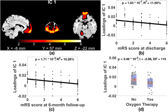
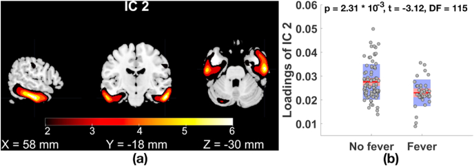
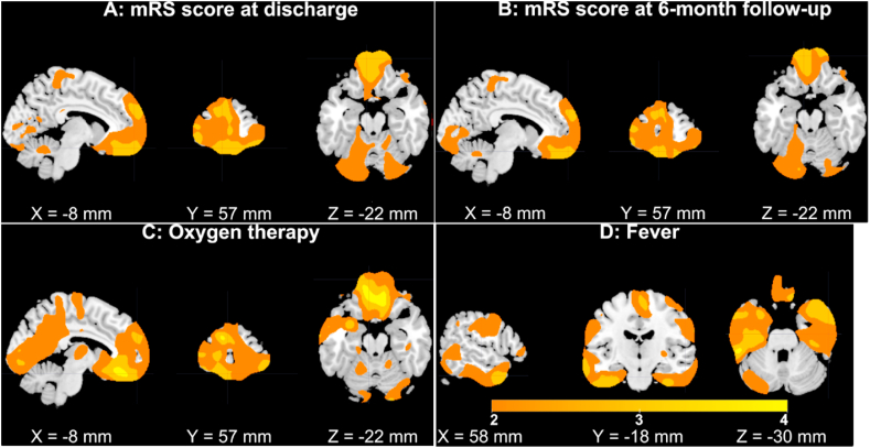
COVID-19, the infectious disease caused by the most recently discovered severe acute respiratory syndrome coronavirus-2 (SARS-CoV-2), has become a global pandemic. It dramatically affects people's health and daily life. Neurological complications are increasingly documented for patients with COVID-19. However, the effect of COVID-19 on the brain is less studied, and existing quantitative neuroimaging analyses of COVID-19 were mainly based on the univariate voxel-based morphometry analysis (VBM) that requires corrections for a large number of tests for statistical significance, multivariate approaches that can reduce the number of tests to be corrected have not been applied to study COVID-19 effect on the brain yet. In this study, we leveraged source-based morphometry (SBM) analysis, a multivariate extension of VBM, to identify changes derived from computed tomography scans in covarying gray matter volume patterns underlying COVID-19 in 120 neurological patients (including 58 cases with COVID-19 and 62 patients without COVID-19 matched for age, gender and diseases). SBM identified that lower gray matter volume (GMV) in superior/medial/middle frontal gyri was significantly associated with a higher level of disability (modified Rankin Scale) at both discharge and six months follow-up phases even when controlling for cerebrovascular diseases. GMV in superior/medial/middle frontal gyri was also significantly reduced in patients receiving oxygen therapy compared to patients not receiving oxygen therapy. Patients with fever presented significant GMV reduction in inferior/middle temporal gyri and fusiform gyrus compared to patients without fever. Patients with agitation showed GMV reduction in superior/medial/middle frontal gyri compared to patients without agitation. Patients with COVID-19 showed no significant GMV differences from patients without COVID-19 in any brain region. Results suggest that COVID-19 may affect the frontal-temporal network in a secondary manner through fever or lack of oxygen.

摘要

新型冠状病毒肺炎(COVID-19)是由最近发现的严重急性呼吸综合征冠状病毒2(SARS-CoV-2)引起的传染病,已成为全球大流行疾病。它极大地影响了人们的健康和日常生活。越来越多的文献记录了COVID-19患者的神经并发症。然而,COVID-19对大脑的影响研究较少,现有的COVID-19定量神经影像学分析主要基于单变量体素形态学分析(VBM),该分析需要对大量测试进行统计显著性校正,而能够减少校正测试数量的多变量方法尚未应用于研究COVID-19对大脑的影响。在本研究中,我们利用基于源的形态学(SBM)分析(VBM的多变量扩展),在120名神经科患者(包括58例COVID-19患者和62例年龄、性别和疾病相匹配的非COVID-19患者)中,识别出COVID-19潜在的共变灰质体积模式中计算机断层扫描产生的变化。SBM发现,即使在控制脑血管疾病的情况下,上/中/额内侧回灰质体积(GMV)较低与出院时和六个月随访期较高的残疾水平(改良Rankin量表)显著相关。与未接受氧疗的患者相比,接受氧疗的患者上/中/额内侧回GMV也显著降低。与无发热患者相比,发热患者颞中/下回和梭状回GMV显著降低。与无躁动患者相比,躁动患者上/中/额内侧回GMV降低。COVID-19患者与非COVID-19患者在任何脑区均未显示出显著的GMV差异。结果表明,COVID-19可能通过发热或缺氧以继发性方式影响额颞叶网络。

https://cdn.ncbi.nlm.nih.gov/pmc/blobs/99e0/8099628/49c3e6615e41/gr4.jpg
https://cdn.ncbi.nlm.nih.gov/pmc/blobs/99e0/8099628/30d695dcbf02/gr1.jpg
https://cdn.ncbi.nlm.nih.gov/pmc/blobs/99e0/8099628/fb99839f87c0/gr2.jpg
https://cdn.ncbi.nlm.nih.gov/pmc/blobs/99e0/8099628/1e21f4c416a0/gr3.jpg
https://cdn.ncbi.nlm.nih.gov/pmc/blobs/99e0/8099628/49c3e6615e41/gr4.jpg
https://cdn.ncbi.nlm.nih.gov/pmc/blobs/99e0/8099628/30d695dcbf02/gr1.jpg
https://cdn.ncbi.nlm.nih.gov/pmc/blobs/99e0/8099628/fb99839f87c0/gr2.jpg
https://cdn.ncbi.nlm.nih.gov/pmc/blobs/99e0/8099628/1e21f4c416a0/gr3.jpg
https://cdn.ncbi.nlm.nih.gov/pmc/blobs/99e0/8099628/49c3e6615e41/gr4.jpg

相似文献

1
Alterations of frontal-temporal gray matter volume associate with clinical measures of older adults with COVID-19.新冠病毒感染老年患者的额颞叶灰质体积改变与临床指标相关。
Neurobiol Stress. 2021 May;14:100326. doi: 10.1016/j.ynstr.2021.100326. Epub 2021 Apr 13.
2
Cortical and Subcortical Gray Matter Volume in Youths With Conduct Problems: A Meta-analysis.有行为问题的青少年的皮质和皮质下灰质体积:一项荟萃分析。
JAMA Psychiatry. 2016 Jan;73(1):64-72. doi: 10.1001/jamapsychiatry.2015.2423.
3
Altered Brain Morphometry in Cerebral Small Vessel Disease With Cerebral Microbleeds: An Investigation Combining Univariate and Multivariate Pattern Analyses.伴有脑微出血的脑小血管病的脑形态测量学改变:一项结合单变量和多变量模式分析的研究
Front Neurol. 2022 Feb 23;13:819055. doi: 10.3389/fneur.2022.819055. eCollection 2022.
4
Decreased gray matter volume and increased white matter volume in patients with neovascular age-related macular degeneration: a voxel-based morphometry study.年龄相关性黄斑变性新生血管患者的灰质体积减少和白质体积增加:基于体素的形态计量学研究。
Aging (Albany NY). 2021 Oct 6;13(19):23182-23192. doi: 10.18632/aging.203610.
5
Decreased gray matter volume is associated with theory of mind deficit in adolescents with schizophrenia.灰质体积减少与精神分裂症青少年的心理理论缺陷有关。
Brain Imaging Behav. 2022 Jun;16(3):1441-1450. doi: 10.1007/s11682-021-00591-9. Epub 2022 Jan 21.
6
White and Gray Matter Volume Changes and Correlation with Visual Evoked Potential in Patients with Optic Neuritis: A Voxel-Based Morphometry Study.视神经炎患者白质和灰质体积变化及其与视觉诱发电位的相关性:基于体素的形态学研究
Med Sci Monit. 2016 Apr 5;22:1115-23. doi: 10.12659/msm.897837.
7
Prognostic Utility of Multivariate Morphometry in Schizophrenia.多变量形态测量学在精神分裂症中的预后效用
Front Psychiatry. 2019 Apr 15;10:245. doi: 10.3389/fpsyt.2019.00245. eCollection 2019.
8
Association Between Diabetes and Gray Matter Atrophy Patterns in a General Older Japanese Population: The Hisayama Study.在一般的老年日本人群中,糖尿病与灰质萎缩模式之间的关联:日山研究。
Diabetes Care. 2022 Jun 2;45(6):1364-1371. doi: 10.2337/dc21-1911.
9
[Voxel-based morphometry study of the brain structures in patients with congenital hereditary hearing loss].先天性遗传性听力损失患者脑结构的基于体素的形态学研究
Zhonghua Er Bi Yan Hou Tou Jing Wai Ke Za Zhi. 2020 Feb 7;55(2):81-86. doi: 10.3760/cma.j.issn.1673-0860.2020.02.001.
10
An investigation of changes in regional gray matter volume in cardiovascular disease patients, pre and post cardiovascular rehabilitation.对心血管疾病患者进行心血管康复治疗前后的区域性灰质体积变化的研究。
Neuroimage Clin. 2013 Oct 6;3:388-95. doi: 10.1016/j.nicl.2013.09.011. eCollection 2013.

引用本文的文献

1
aNy-way ICA and its application to estimate cortico-thalamo-cerebellar functional links in schizophrenia.无论如何,独立成分分析(ICA)及其在估计精神分裂症患者皮质-丘脑-小脑功能连接中的应用。
bioRxiv. 2025 Jun 6:2025.06.02.657541. doi: 10.1101/2025.06.02.657541.
2
Longitudinal alterations in morphological brain networks and cognitive function in common-type COVID-19: a 3-month follow-up study.普通型新冠病毒肺炎患者脑形态学网络和认知功能的纵向变化:一项为期3个月的随访研究
Front Neurol. 2025 Apr 15;16:1549195. doi: 10.3389/fneur.2025.1549195. eCollection 2025.
3
Diffusion tensor imaging features of white matter pathways in the brain after COVID-19 infection.

本文引用的文献

1
Brain volumetric changes in the general population following the COVID-19 outbreak and lockdown.新冠疫情爆发及封控措施后普通人群的脑容量变化。
Neuroimage. 2021 Oct 1;239:118311. doi: 10.1016/j.neuroimage.2021.118311. Epub 2021 Jun 26.
2
F-FDG brain PET hypometabolism in patients with long COVID.长新冠患者的 F-FDG 脑 PET 代谢低下。
Eur J Nucl Med Mol Imaging. 2021 Aug;48(9):2823-2833. doi: 10.1007/s00259-021-05215-4. Epub 2021 Jan 26.
3
Neuroinvasion and Encephalitis Following Intranasal Inoculation of SARS-CoV-2 in K18-hACE2 Mice.
新型冠状病毒肺炎感染后脑白质通路的扩散张量成像特征
Radiologie (Heidelb). 2025 Jan 28. doi: 10.1007/s00117-024-01414-w.
4
Structural MRI correlates of cognitive and neuropsychiatric symptoms in Long COVID: a pilot study.新冠后综合征中认知和神经精神症状的结构磁共振成像相关性:一项试点研究
Front Psychiatry. 2024 Dec 4;15:1412020. doi: 10.3389/fpsyt.2024.1412020. eCollection 2024.
5
Acute and long-term effects of COVID-19 on brain and mental health: A narrative review.新型冠状病毒肺炎对大脑和心理健康的急性及长期影响:一项叙述性综述
Brain Behav Immun. 2025 Jan;123:928-945. doi: 10.1016/j.bbi.2024.11.007. Epub 2024 Nov 3.
6
COVID-19 related cognitive, structural and functional brain changes among Italian adolescents and young adults: a multimodal longitudinal case-control study.意大利青少年和青年中与新冠病毒病相关的认知、结构和功能性脑变化:一项多模态纵向病例对照研究
Transl Psychiatry. 2024 Oct 2;14(1):402. doi: 10.1038/s41398-024-03108-2.
7
Altered functional brain connectivity, efficiency, and information flow associated with brain fog after mild to moderate COVID-19 infection.轻度至中度 COVID-19 感染后与脑雾相关的大脑功能连接、效率和信息流的改变。
Sci Rep. 2024 Sep 27;14(1):22094. doi: 10.1038/s41598-024-73311-0.
8
Tracts in the limbic system show microstructural alterations post COVID-19 recovery.在新冠病毒感染康复后,边缘系统中的神经束显示出微观结构改变。
Brain Commun. 2024 May 7;6(3):fcae139. doi: 10.1093/braincomms/fcae139. eCollection 2024.
9
Searching Reproducible Brain Features using NeuroMark: Templates for Different Age Populations and Imaging Modalities.使用NeuroMark搜索可重现的脑特征:针对不同年龄人群和成像模态的模板
Neuroimage. 2024 Apr 15;292:120617. doi: 10.1016/j.neuroimage.2024.120617. Epub 2024 Apr 16.
10
Brain temperature and free water increases after mild COVID-19 infection.轻度 COVID-19 感染后大脑温度和自由水增加。
Sci Rep. 2024 Mar 28;14(1):7450. doi: 10.1038/s41598-024-57561-6.
经鼻腔接种 SARS-CoV-2 后 K18-hACE2 小鼠的神经侵袭和脑炎。
Viruses. 2021 Jan 19;13(1):132. doi: 10.3390/v13010132.
4
The cerebral network of COVID-19-related encephalopathy: a longitudinal voxel-based 18F-FDG-PET study.新型冠状病毒肺炎相关脑病的脑网络:一项基于体素的18F-氟代脱氧葡萄糖正电子发射断层扫描纵向研究
Eur J Nucl Med Mol Imaging. 2021 Jul;48(8):2543-2557. doi: 10.1007/s00259-020-05178-y. Epub 2021 Jan 15.
5
Neuroimaging findings of brain MRI and CT in patients with COVID-19: A systematic review and meta-analysis.新型冠状病毒肺炎患者脑 MRI 和 CT 的神经影像学表现:系统评价和荟萃分析。
Eur J Radiol. 2020 Dec;133:109393. doi: 10.1016/j.ejrad.2020.109393. Epub 2020 Nov 3.
6
Posterior Reversible Encephalopathy Syndrome in Patients with Coronavirus Disease 2019: Two Cases and A Review of The Literature.新型冠状病毒 2019 患者的后部可逆性脑病综合征:两例病例及文献复习。
J Stroke Cerebrovasc Dis. 2020 Nov;29(11):105212. doi: 10.1016/j.jstrokecerebrovasdis.2020.105212. Epub 2020 Jul 30.
7
COVID-19 impact on consecutive neurological patients admitted to the emergency department.新型冠状病毒肺炎对急诊科连续收治的神经系统疾病患者的影响。
J Neurol Neurosurg Psychiatry. 2021 Feb;92(2):218-220. doi: 10.1136/jnnp-2020-323929. Epub 2020 Oct 14.
8
A Prospective Study of Neurologic Disorders in Hospitalized Patients With COVID-19 in New York City.一项关于纽约市住院 COVID-19 患者神经系统疾病的前瞻性研究。
Neurology. 2021 Jan 26;96(4):e575-e586. doi: 10.1212/WNL.0000000000010979. Epub 2020 Oct 5.
9
Fever Patterns, Cytokine Profiles, and Outcomes in COVID-19.新型冠状病毒肺炎的发热模式、细胞因子谱及预后
Open Forum Infect Dis. 2020 Aug 24;7(9):ofaa375. doi: 10.1093/ofid/ofaa375. eCollection 2020 Sep.
10
Clinical Presentation and Outcomes of Severe Acute Respiratory Syndrome Coronavirus 2-Related Encephalitis: The ENCOVID Multicenter Study.严重急性呼吸综合征冠状病毒 2 相关脑炎的临床特征和结局:ENCOVID 多中心研究。
J Infect Dis. 2021 Jan 4;223(1):28-37. doi: 10.1093/infdis/jiaa609.