Mathematical Neuro-Oncology Lab, Precision Neurotherapeutics Innovation Program, Department of Neurological Surgery, Mayo Clinic, 5777 E Mayo Blvd, Phoenix, AZ, 85054, USA.
Mayo Clinic School of Medicine, Rochester, MN, USA.
Sci Rep. 2021 Dec 1;11(1):23202. doi: 10.1038/s41598-021-02495-6.
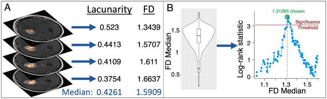
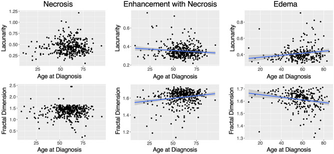
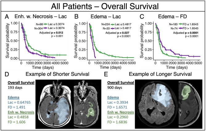
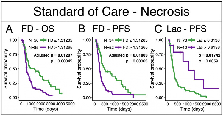
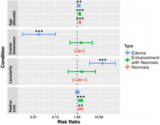
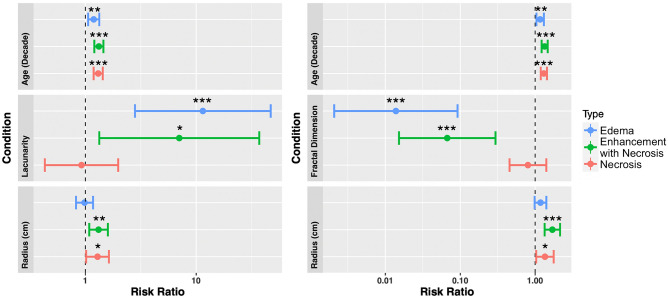
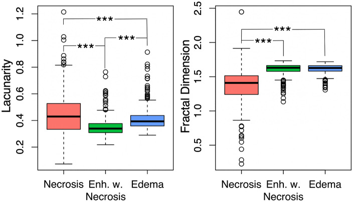
Lacunarity, a quantitative morphological measure of how shapes fill space, and fractal dimension, a morphological measure of the complexity of pixel arrangement, have shown relationships with outcome across a variety of cancers. However, the application of these metrics to glioblastoma (GBM), a very aggressive primary brain tumor, has not been fully explored. In this project, we computed lacunarity and fractal dimension values for GBM-induced abnormalities on clinically standard magnetic resonance imaging (MRI). In our patient cohort (n = 402), we connect these morphological metrics calculated on pretreatment MRI with the survival of patients with GBM. We calculated lacunarity and fractal dimension on necrotic regions (n = 390), all abnormalities present on T1Gd MRI (n = 402), and abnormalities present on T2/FLAIR MRI (n = 257). We also explored the relationship between these metrics and age at diagnosis, as well as abnormality volume. We found statistically significant relationships to outcome for all three imaging regions that we tested, with the shape of T2/FLAIR abnormalities that are typically associated with edema showing the strongest relationship with overall survival. This link between morphological and survival metrics could be driven by underlying biological phenomena, tumor location or microenvironmental factors that should be further explored.
分形维数和空隙度是用于衡量形状填充空间的定量形态学参数,以及用于衡量像素排列复杂性的形态学参数,它们与多种癌症的预后结果均具有相关性。然而,这些指标在胶质母细胞瘤(GBM)这种非常侵袭性的原发性脑肿瘤中的应用尚未得到充分探索。在本项目中,我们计算了 GBM 诱导的异常在临床标准磁共振成像(MRI)上的分形维数和空隙度值。在我们的患者队列中(n=402),我们将这些在预处理 MRI 上计算出的形态学指标与 GBM 患者的生存情况联系起来。我们分别计算了坏死区域(n=390)、T1Gd MRI 上所有异常(n=402)以及 T2/FLAIR MRI 上异常(n=257)的分形维数和空隙度。我们还探索了这些指标与诊断时年龄以及异常体积之间的关系。我们发现,在所测试的所有三个成像区域中,这些指标与预后结果均具有统计学意义的相关性,而与水肿相关的 T2/FLAIR 异常的形状与总生存率之间的相关性最强。这种形态学和生存指标之间的联系可能是由潜在的生物学现象、肿瘤位置或微环境因素所驱动的,这些因素需要进一步探索。