• 文献检索
  • 文档翻译
  • 深度研究
  • 学术资讯
  • Suppr Zotero 插件Zotero 插件
  • 邀请有礼
  • 套餐&价格
  • 历史记录
应用&插件
Suppr Zotero 插件Zotero 插件浏览器插件Mac 客户端Windows 客户端微信小程序
定价
高级版会员购买积分包购买API积分包
服务
文献检索文档翻译深度研究API 文档MCP 服务
关于我们
关于 Suppr公司介绍联系我们用户协议隐私条款
关注我们

Suppr 超能文献

核心技术专利:CN118964589B侵权必究
粤ICP备2023148730 号-1Suppr @ 2026

文献检索

告别复杂PubMed语法,用中文像聊天一样搜索,搜遍4000万医学文献。AI智能推荐,让科研检索更轻松。

立即免费搜索

文件翻译

保留排版,准确专业,支持PDF/Word/PPT等文件格式,支持 12+语言互译。

免费翻译文档

深度研究

AI帮你快速写综述,25分钟生成高质量综述,智能提取关键信息,辅助科研写作。

立即免费体验

具有可解释性的脑磁共振成像自动病变和特征提取管道

Automated Lesion and Feature Extraction Pipeline for Brain MRIs with Interpretability.

作者信息

Eghbali Reza, Nedelec Pierre, Weiss David, Bhalerao Radhika, Xie Long, Rudie Jeffrey D, Liu Chunlei, Sugrue Leo P, Rauschecker Andreas M

机构信息

Department of Radiology and Biomedical Imaging, University of California, San Francisco, San Francisco, CA, USA.

Berkeley Institute for Data Science, University of California, Berkeley, Berkeley, CA, USA.

出版信息

Neuroinformatics. 2025 Jan 9;23(1):2. doi: 10.1007/s12021-024-09708-z.

DOI:10.1007/s12021-024-09708-z
PMID:39786657
原文链接:https://pmc.ncbi.nlm.nih.gov/articles/PMC11717894/
Abstract

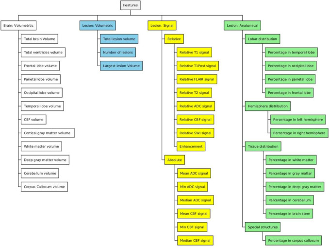
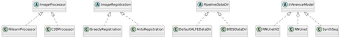
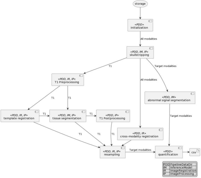
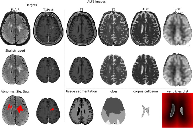
This paper introduces the Automated Lesion and Feature Extraction (ALFE) pipeline, an open-source, Python-based pipeline that consumes MR images of the brain and produces anatomical segmentations, lesion segmentations, and human-interpretable imaging features describing the lesions in the brain. ALFE pipeline is modeled after the neuroradiology workflow and generates features that can be used by physicians for quantitative analysis of clinical brain MRIs and for machine learning applications. The pipeline uses a decoupled design which allows the user to customize the image processing, image registrations, and AI segmentation tools without the need to change the business logic of the pipeline. In this manuscript, we give an overview of ALFE, present the main aspects of ALFE pipeline design philosophy, and present case studies.

摘要

本文介绍了自动病变与特征提取(ALFE)流程,这是一个基于Python的开源流程,它接收脑部的磁共振成像(MR)图像,并生成解剖分割、病变分割以及描述脑部病变的可被人类解读的成像特征。ALFE流程是仿照神经放射学工作流程建模的,生成的特征可供医生用于临床脑部MRI的定量分析以及机器学习应用。该流程采用解耦设计,允许用户定制图像处理、图像配准和人工智能分割工具,而无需更改流程的业务逻辑。在本手稿中,我们对ALFE进行了概述,介绍了ALFE流程设计理念的主要方面,并展示了案例研究。

https://cdn.ncbi.nlm.nih.gov/pmc/blobs/70dd/11717894/da0857ef98a4/12021_2024_9708_Fig6_HTML.jpg
https://cdn.ncbi.nlm.nih.gov/pmc/blobs/70dd/11717894/3bea9d75d362/12021_2024_9708_Fig1_HTML.jpg
https://cdn.ncbi.nlm.nih.gov/pmc/blobs/70dd/11717894/735e16dad8c7/12021_2024_9708_Fig2_HTML.jpg
https://cdn.ncbi.nlm.nih.gov/pmc/blobs/70dd/11717894/015e36bd8b2f/12021_2024_9708_Fig3_HTML.jpg
https://cdn.ncbi.nlm.nih.gov/pmc/blobs/70dd/11717894/239b79cfa136/12021_2024_9708_Fig4_HTML.jpg
https://cdn.ncbi.nlm.nih.gov/pmc/blobs/70dd/11717894/78193be8c22d/12021_2024_9708_Fig5_HTML.jpg
https://cdn.ncbi.nlm.nih.gov/pmc/blobs/70dd/11717894/da0857ef98a4/12021_2024_9708_Fig6_HTML.jpg

相似文献

1
Automated Lesion and Feature Extraction Pipeline for Brain MRIs with Interpretability.具有可解释性的脑磁共振成像自动病变和特征提取管道
Neuroinformatics. 2025 Jan 9;23(1):2. doi: 10.1007/s12021-024-09708-z.
2
Improving brain atrophy quantification with deep learning from automated labels using tissue similarity priors.利用组织相似性先验从自动标签中通过深度学习改善脑萎缩定量。
Comput Biol Med. 2024 Sep;179:108811. doi: 10.1016/j.compbiomed.2024.108811. Epub 2024 Jul 10.
3
Brain tumor segmentation and detection in MRI using convolutional neural networks and VGG16.使用卷积神经网络和VGG16在磁共振成像(MRI)中进行脑肿瘤分割与检测
Cancer Biomark. 2025 Mar;42(3):18758592241311184. doi: 10.1177/18758592241311184. Epub 2025 Apr 4.
4
SEPIA-Susceptibility mapping pipeline tool for phase images.SEPIA 相位图像磁化率映射分析流水线工具
Neuroimage. 2021 Feb 15;227:117611. doi: 10.1016/j.neuroimage.2020.117611. Epub 2020 Dec 10.
5
Convolutional Neural Network for Automated FLAIR Lesion Segmentation on Clinical Brain MR Imaging.基于卷积神经网络的临床脑部磁共振成像 FLAIR 病变分割自动化。
AJNR Am J Neuroradiol. 2019 Aug;40(8):1282-1290. doi: 10.3174/ajnr.A6138. Epub 2019 Jul 25.
6
A multi-atlas based method for automated anatomical rat brain MRI segmentation and extraction of PET activity.一种基于多图谱的自动大鼠脑解剖MRI分割及PET活性提取方法。
PLoS One. 2014 Oct 17;9(10):e109113. doi: 10.1371/journal.pone.0109113. eCollection 2014.
7
A generalizable brain extraction net (BEN) for multimodal MRI data from rodents, nonhuman primates, and humans.一种可推广的用于啮齿动物、非人灵长类动物和人类多模态 MRI 数据的大脑提取网络(BEN)。
Elife. 2022 Dec 22;11:e81217. doi: 10.7554/eLife.81217.
8
Infant FreeSurfer: An automated segmentation and surface extraction pipeline for T1-weighted neuroimaging data of infants 0-2 years.婴儿 FreeSurfer:用于 0-2 岁婴儿 T1 加权神经影像学数据的自动分割和表面提取管道。
Neuroimage. 2020 Sep;218:116946. doi: 10.1016/j.neuroimage.2020.116946. Epub 2020 May 20.
9
Task-based assessment of a convolutional neural network for segmenting breast lesions for radiomic analysis.基于任务的卷积神经网络在乳腺病变分割中的放射组学分析评估。
Magn Reson Med. 2019 Aug;82(2):786-795. doi: 10.1002/mrm.27758. Epub 2019 Apr 8.
10
Whole volume brain extraction for multi-centre, multi-disease FLAIR MRI datasets.用于多中心、多疾病液体衰减反转恢复(FLAIR)磁共振成像(MRI)数据集的全脑体积提取
Magn Reson Imaging. 2020 Feb;66:116-130. doi: 10.1016/j.mri.2019.08.022. Epub 2019 Aug 28.

本文引用的文献

1
The University of California San Francisco Brain Metastases Stereotactic Radiosurgery (UCSF-BMSR) MRI Dataset.加利福尼亚大学旧金山分校脑转移瘤立体定向放射外科手术(UCSF - BMSR)MRI数据集
Radiol Artif Intell. 2024 Mar;6(2):e230126. doi: 10.1148/ryai.230126.
2
Evaluating Autoencoders for Dimensionality Reduction of MRI-derived Radiomics and Classification of Malignant Brain Tumors.评估自动编码器在降低MRI衍生的放射组学维度及恶性脑肿瘤分类中的应用
Sci Stat Database Manag. 2023 Jul;2023. doi: 10.1145/3603719.3603737. Epub 2023 Aug 27.
3
Radiomics-Based Machine Learning Model for Predicting Overall and Progression-Free Survival in Rare Cancer: A Case Study for Primary CNS Lymphoma Patients.
基于影像组学的机器学习模型预测罕见癌症的总生存期和无进展生存期:原发性中枢神经系统淋巴瘤患者的案例研究
Bioengineering (Basel). 2023 Feb 22;10(3):285. doi: 10.3390/bioengineering10030285.
4
SynthSeg: Segmentation of brain MRI scans of any contrast and resolution without retraining.SynthSeg:无需重新训练即可对任何对比度和分辨率的脑 MRI 扫描进行分割。
Med Image Anal. 2023 May;86:102789. doi: 10.1016/j.media.2023.102789. Epub 2023 Feb 25.
5
Combining radiomics and deep convolutional neural network features from preoperative MRI for predicting clinically relevant genetic biomarkers in glioblastoma.结合术前磁共振成像的影像组学和深度卷积神经网络特征以预测胶质母细胞瘤中临床相关的遗传生物标志物。
Neurooncol Adv. 2022 Apr 22;4(1):vdac060. doi: 10.1093/noajnl/vdac060. eCollection 2022 Jan-Dec.
6
Clinical measures, radiomics, and genomics offer synergistic value in AI-based prediction of overall survival in patients with glioblastoma.临床指标、影像组学和基因组学为基于人工智能的胶质母细胞瘤患者总生存期预测提供了协同价值。
Sci Rep. 2022 May 24;12(1):8784. doi: 10.1038/s41598-022-12699-z.
7
Shape matters: morphological metrics of glioblastoma imaging abnormalities as biomarkers of prognosis.形态很重要:胶质母细胞瘤影像学异常的形态学指标可作为预后的生物标志物。
Sci Rep. 2021 Dec 1;11(1):23202. doi: 10.1038/s41598-021-02495-6.
8
Automated multiclass tissue segmentation of clinical brain MRIs with lesions.基于临床脑 MRI 的病变的自动多类组织分割。
Neuroimage Clin. 2021;31:102769. doi: 10.1016/j.nicl.2021.102769. Epub 2021 Jul 24.
9
Subspecialty-Level Deep Gray Matter Differential Diagnoses with Deep Learning and Bayesian Networks on Clinical Brain MRI: A Pilot Study.基于临床脑磁共振成像的深度学习和贝叶斯网络进行亚专业水平深部灰质鉴别诊断:一项初步研究
Radiol Artif Intell. 2020 Sep 23;2(5):e190146. doi: 10.1148/ryai.2020190146. eCollection 2020 Sep.
10
nnU-Net: a self-configuring method for deep learning-based biomedical image segmentation.nnU-Net:一种基于深度学习的生物医学图像分割的自配置方法。
Nat Methods. 2021 Feb;18(2):203-211. doi: 10.1038/s41592-020-01008-z. Epub 2020 Dec 7.