• 文献检索
  • 文档翻译
  • 深度研究
  • 学术资讯
  • Suppr Zotero 插件Zotero 插件
  • 邀请有礼
  • 套餐&价格
  • 历史记录
应用&插件
Suppr Zotero 插件Zotero 插件浏览器插件Mac 客户端Windows 客户端微信小程序
定价
高级版会员购买积分包购买API积分包
服务
文献检索文档翻译深度研究API 文档MCP 服务
关于我们
关于 Suppr公司介绍联系我们用户协议隐私条款
关注我们

Suppr 超能文献

核心技术专利:CN118964589B侵权必究
粤ICP备2023148730 号-1Suppr @ 2026

文献检索

告别复杂PubMed语法,用中文像聊天一样搜索,搜遍4000万医学文献。AI智能推荐,让科研检索更轻松。

立即免费搜索

文件翻译

保留排版,准确专业,支持PDF/Word/PPT等文件格式,支持 12+语言互译。

免费翻译文档

深度研究

AI帮你快速写综述,25分钟生成高质量综述,智能提取关键信息,辅助科研写作。

立即免费体验

泡球蚴原头节与人肝星状细胞共培养中肝纤维化相关蛋白的结构变化和表达。

Structural changes and expression of hepatic fibrosis-related proteins in coculture of Echinococcus multilocularis protoscoleces and human hepatic stellate cells.

机构信息

Department of Human Parasitology, Guilin Medical University, Guilin, 541199, Guangxi Zhuang, China.

Department of Immunology, Faculty of Medicine, Qinghai University, Xining, 810001, Qinghai, China.

出版信息

Parasit Vectors. 2021 Dec 2;14(1):593. doi: 10.1186/s13071-021-05037-1.

DOI:10.1186/s13071-021-05037-1
PMID:34857049
原文链接:https://pmc.ncbi.nlm.nih.gov/articles/PMC8641223/
Abstract

BACKGROUND

Echinococcus multilocularis is the causative agent of human hepatic alveolar echinococcosis (AE). AE can cause damage to several organs, primarily the liver, and have severe outcomes, such as hepatic failure and encephalopathy. The main purpose of this study was to explore the interactions between hepatic stellate cells (HSCs) and E. multilocularis protoscoleces (PSCs). The results of this study provide an experimental basis for further examination of the pathogenesis of hepatic fibrosis due to AE infection.

METHODS

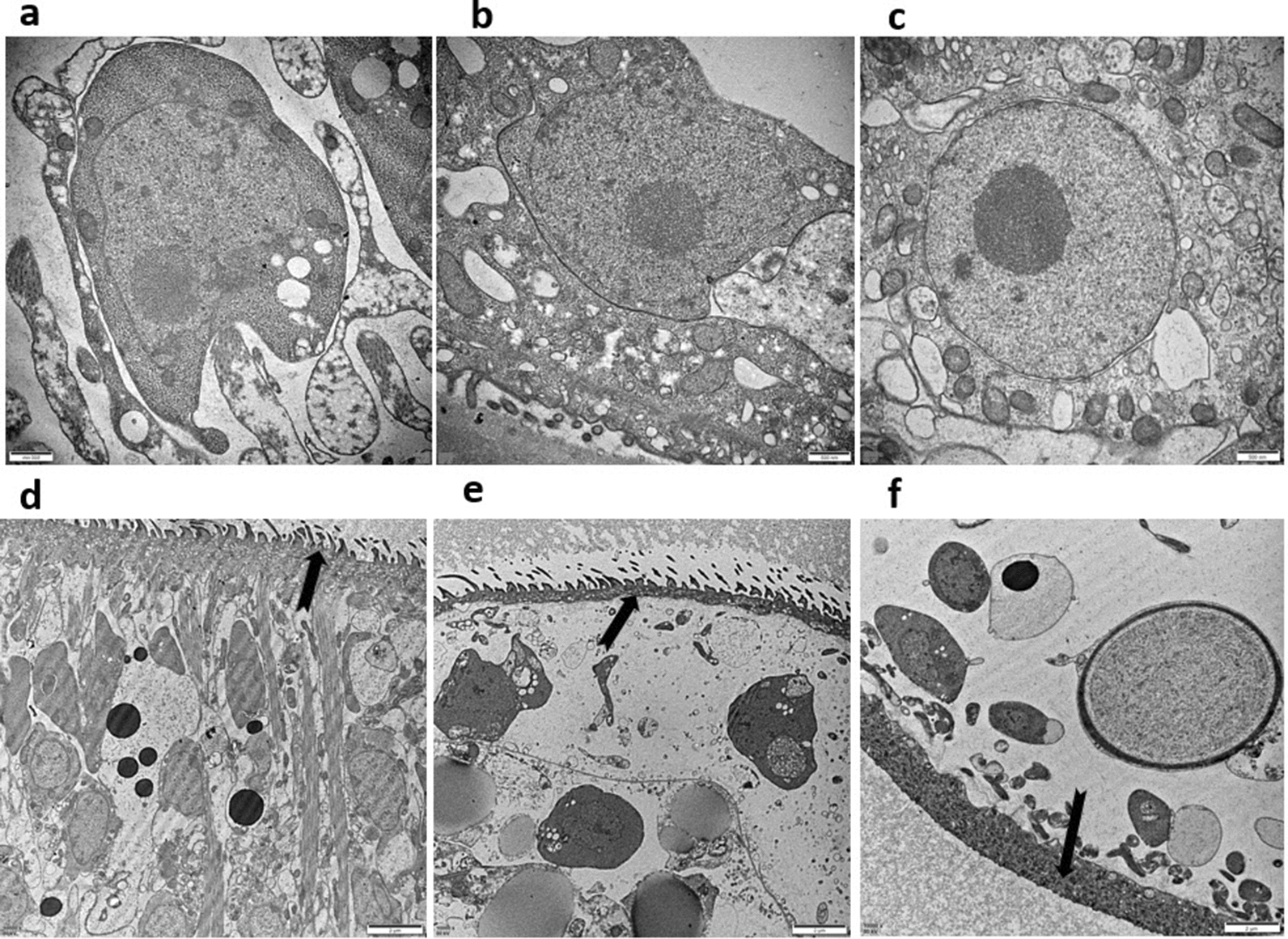
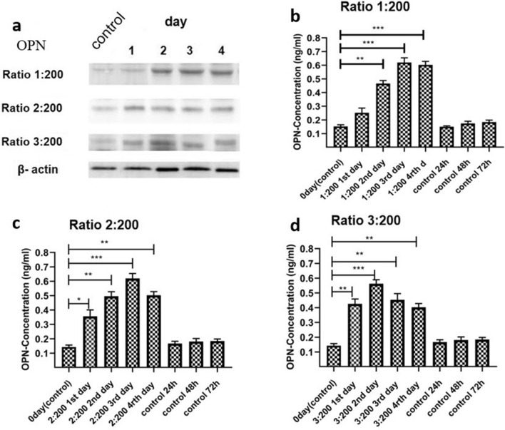
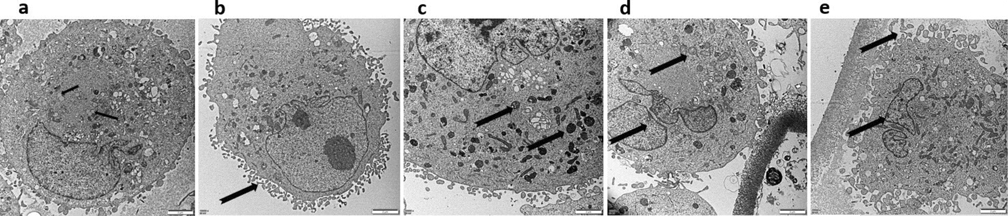
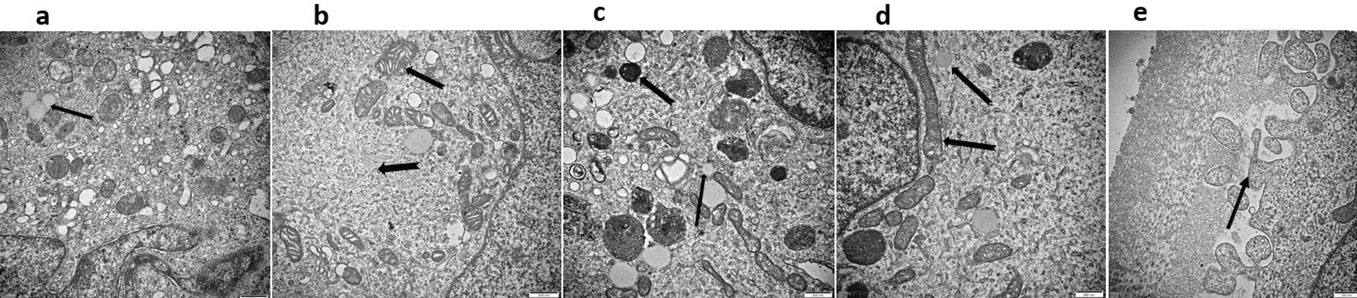
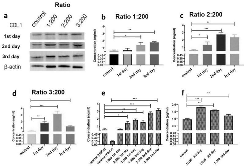
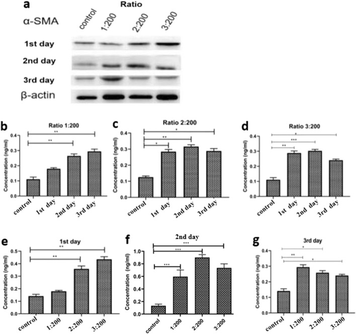
We investigated the role of Echinococcus multilocularis (Echinococcus genus) PSCs in hepatic fibrosis by examining structural changes and measuring hepatic fibrosis-related protein levels in cocultures of PSCs and human HSCs. Structural changes were detected by transmission electron microscopy (TEM), and levels of the hepatic fibrosis-related proteins collagen I (Col-I), alpha-smooth muscle actin (α-SMA) and osteopontin (OPN) were measured by western blotting and enzyme-linked immunosorbent assay (ELISA).

RESULTS

Under coculture (1) both PSCs and HSCs exhibited morphological changes, as observed by TEM; (2) Col-I, α-SMA, and OPN expression levels, which were determined by western blotting and ELISA, significantly increased after 3 days of incubation.

CONCLUSIONS

The results of this study provide insights into the molecular mechanisms of AE-induced hepatic fibrosis.

摘要

背景

多房棘球绦虫是人体泡型包虫病(AE)的病原体。AE 可导致多个器官受损,主要是肝脏,并导致严重后果,如肝衰竭和脑病。本研究的主要目的是探讨肝星状细胞(HSCs)与多房棘球蚴原头节(PSCs)之间的相互作用。本研究的结果为进一步研究 AE 感染引起的肝纤维化发病机制提供了实验依据。

方法

通过观察 PSCs 和人 HSCs 共培养物中的结构变化和测量肝纤维化相关蛋白水平,研究多房棘球绦虫(棘球属)PSCs 在肝纤维化中的作用。通过透射电子显微镜(TEM)检测结构变化,通过 Western blot 和酶联免疫吸附试验(ELISA)测量肝纤维化相关蛋白胶原 I(Col-I)、α-平滑肌肌动蛋白(α-SMA)和骨桥蛋白(OPN)的水平。

结果

共培养(1)PSCs 和 HSCs 均发生形态学变化,TEM 观察到;(2)孵育 3 天后,Western blot 和 ELISA 测定 Col-I、α-SMA 和 OPN 的表达水平显著增加。

结论

本研究结果为了解 AE 诱导的肝纤维化的分子机制提供了线索。

https://cdn.ncbi.nlm.nih.gov/pmc/blobs/089f/8641223/df96017633b0/13071_2021_5037_Fig6_HTML.jpg
https://cdn.ncbi.nlm.nih.gov/pmc/blobs/089f/8641223/75bdb9d0088f/13071_2021_5037_Fig1_HTML.jpg
https://cdn.ncbi.nlm.nih.gov/pmc/blobs/089f/8641223/4227c1f32a2b/13071_2021_5037_Fig2_HTML.jpg
https://cdn.ncbi.nlm.nih.gov/pmc/blobs/089f/8641223/dac9b71f8ab3/13071_2021_5037_Fig3_HTML.jpg
https://cdn.ncbi.nlm.nih.gov/pmc/blobs/089f/8641223/974a9e99b1d0/13071_2021_5037_Fig4_HTML.jpg
https://cdn.ncbi.nlm.nih.gov/pmc/blobs/089f/8641223/ac41fd503835/13071_2021_5037_Fig5_HTML.jpg
https://cdn.ncbi.nlm.nih.gov/pmc/blobs/089f/8641223/df96017633b0/13071_2021_5037_Fig6_HTML.jpg

相似文献

1
Structural changes and expression of hepatic fibrosis-related proteins in coculture of Echinococcus multilocularis protoscoleces and human hepatic stellate cells.泡球蚴原头节与人肝星状细胞共培养中肝纤维化相关蛋白的结构变化和表达。
Parasit Vectors. 2021 Dec 2;14(1):593. doi: 10.1186/s13071-021-05037-1.
2
[Effects of persistent infections on hepatic fibrosis in mice].[持续性感染对小鼠肝纤维化的影响]
Zhongguo Xue Xi Chong Bing Fang Zhi Za Zhi. 2021 Feb 3;33(1):54-61. doi: 10.16250/j.32.1374.2020282.
3
An improved experimental method for simultaneously isolating hepatocytes and hepatic stellate cells in mouse liver infected with Echinococcus multilocularis.一种改良的实验方法,用于同时分离感染多房棘球蚴的小鼠肝脏中的肝细胞和肝星状细胞。
J Clin Lab Anal. 2021 Dec;35(12):e24084. doi: 10.1002/jcla.24084. Epub 2021 Nov 1.
4
[The correlation between osteopontin and metastasis of hepatic Echinococcus multilocularis infection].[骨桥蛋白与肝泡状棘球蚴感染转移的相关性]
Zhongguo Ji Sheng Chong Xue Yu Ji Sheng Chong Bing Za Zhi. 2011 Feb 28;29(1):33-6.
5
[Dynamic observation on capillarization of liver sinusoidal endothelial cells induced by infection].[感染诱导肝窦内皮细胞毛细血管化的动态观察]
Zhongguo Xue Xi Chong Bing Fang Zhi Za Zhi. 2024 Apr 1;36(1):34-43. doi: 10.16250/j.32.1374.2023243.
6
drives the polarization of macrophages by regulating the RhoA-MAPK signaling pathway and thus affects liver fibrosis.通过调节 RhoA-MAPK 信号通路来驱动巨噬细胞的极化,从而影响肝纤维化。
Bioengineered. 2022 Apr;13(4):8747-8758. doi: 10.1080/21655979.2022.2056690.
7
Osteopontin promotes collagen I synthesis in hepatic stellate cells by miRNA-129-5p inhibition.骨桥蛋白通过 miRNA-129-5p 抑制促进肝星状细胞胶原 I 的合成。
Exp Cell Res. 2018 Jan 15;362(2):343-348. doi: 10.1016/j.yexcr.2017.11.035. Epub 2017 Dec 2.
8
[Comparison observation on the mature alveolar of Echinococcus sibiricensis and Echinococcus multilocularis in the experimentally infected white mice].[实验感染小白鼠中细粒棘球绦虫和多房棘球绦虫成熟泡球蚴的比较观察]
Shi Yan Sheng Wu Xue Bao. 2001 Dec;34(4):261-8.
9
TGF-β and TGF-β/Smad signaling in the interactions between Echinococcus multilocularis and its hosts.棘球蚴绦虫与其宿主相互作用中的 TGF-β 和 TGF-β/Smad 信号通路。
PLoS One. 2013;8(2):e55379. doi: 10.1371/journal.pone.0055379. Epub 2013 Feb 6.
10
Autocrine osteopontin is involved in maintaining the growth and metastasis of Echinococcus multilocularis.自分泌骨桥蛋白参与多房棘球蚴的生长和转移。
Acta Trop. 2022 Apr;228:106328. doi: 10.1016/j.actatropica.2022.106328. Epub 2022 Jan 24.

引用本文的文献

1
Preliminary Proteomic and Metabolomic Analyses Reveal Potential Serum Biomarkers for Identifying Alveolar Echinococcosis in Mice.初步蛋白质组学和代谢组学分析揭示了用于鉴定小鼠肺泡型棘球蚴病的潜在血清生物标志物。
Vet Sci. 2025 Jun 9;12(6):565. doi: 10.3390/vetsci12060565.
2
Activation of the Wnt signaling pathway and its role in epithelial-mesenchymal transition and hepatic fibrosis in alveolar echinococcosis.Wnt信号通路的激活及其在泡型棘球蚴病上皮-间质转化和肝纤维化中的作用
Front Cell Infect Microbiol. 2025 May 27;15:1583802. doi: 10.3389/fcimb.2025.1583802. eCollection 2025.
3
-Induced Liver Damage Through Ferroptosis in Rat Model.

本文引用的文献

1
[Effects of persistent infections on hepatic fibrosis in mice].[持续性感染对小鼠肝纤维化的影响]
Zhongguo Xue Xi Chong Bing Fang Zhi Za Zhi. 2021 Feb 3;33(1):54-61. doi: 10.16250/j.32.1374.2020282.
2
In vitro metabolomic footprint of the Echinococcus multilocularis metacestode.体外代谢组学对细粒棘球蚴原头蚴的研究。
Sci Rep. 2019 Dec 19;9(1):19438. doi: 10.1038/s41598-019-56073-y.
3
Mechanism of Fibrosis Induced by spp.由……物种诱导的纤维化机制
- 大鼠模型中通过铁死亡诱导的肝损伤。
Cells. 2025 Feb 22;14(5):328. doi: 10.3390/cells14050328.
Diseases. 2019 Aug 12;7(3):51. doi: 10.3390/diseases7030051.
4
The role of fibroblast growth factor signalling in Echinococcus multilocularis development and host-parasite interaction.成纤维细胞生长因子信号通路在泡球蚴发育和宿主-寄生虫相互作用中的作用。
PLoS Negl Trop Dis. 2019 Mar 8;13(3):e0006959. doi: 10.1371/journal.pntd.0006959. eCollection 2019 Mar.
5
Echinococcus granulosus cyst fluid enhances epithelial-mesenchymal transition.细粒棘球绦虫囊液可增强上皮-间质转化。
Parasite Immunol. 2018 Jun;40(6):e12533. doi: 10.1111/pim.12533.
6
Three-dimensional hepatocyte culture system for the study of Echinococcus multilocularis larval development.用于研究细粒棘球蚴幼虫发育的三维肝细胞培养系统。
PLoS Negl Trop Dis. 2018 Mar 14;12(3):e0006309. doi: 10.1371/journal.pntd.0006309. eCollection 2018 Mar.
7
In vivo treatment with IL-17A attenuates hydatid cyst growth and liver fibrogenesis in an experimental model of echinococcosis.在棘球蚴病实验模型中,用白细胞介素-17A进行体内治疗可减弱包虫囊肿生长和肝纤维化。
Acta Trop. 2018 May;181:6-10. doi: 10.1016/j.actatropica.2018.01.014. Epub 2018 Feb 3.
8
Echinococcus-Host Interactions at Cellular and Molecular Levels.细胞和分子水平上的棘球蚴与宿主的相互作用
Adv Parasitol. 2017;95:147-212. doi: 10.1016/bs.apar.2016.09.001. Epub 2016 Nov 21.
9
Liver fibrosis and hepatic stellate cells: Etiology, pathological hallmarks and therapeutic targets.肝纤维化与肝星状细胞:病因、病理特征及治疗靶点
World J Gastroenterol. 2016 Dec 28;22(48):10512-10522. doi: 10.3748/wjg.v22.i48.10512.
10
In vitro culture of Echinococcus multilocularis producing protoscoleces and mouse infection with the cultured vesicles.体外培养多房棘球绦虫产生原头蚴,并感染培养囊泡的小鼠。
Parasit Vectors. 2016 Jul 25;9(1):411. doi: 10.1186/s13071-016-1687-y.