Visual Physiology, School of Allied Health Sciences, Kitasato University, Kanagawa, Japan.
Department of Ophthalmology, School of Medicine, Kitasato University, Kanagawa, Japan.
Biomed Res Int. 2021 Nov 27;2021:5752248. doi: 10.1155/2021/5752248. eCollection 2021.
To assess the effect of platelet-rich plasma (PRP) on the healing response of the corneal epithelium in eyes undergoing phototherapeutic keratectomy (PTK).
We prospectively examined 20 eyes of 10 patients undergoing bilateral PTK for granular corneal dystrophy or band keratopathy. Patients were randomly assigned to start topical administration of PRP ophthalmic suspension (PRP group) or artificial tears (control group) 4 times daily for 2 weeks. Immediately, 1, and 2 days, and 1 week after PTK, we quantitatively measured the staining area of the corneal epithelium, using slit-lamp photography. We also determined the subjective symptoms and the satisfaction, using the visual analogue system (VAS).
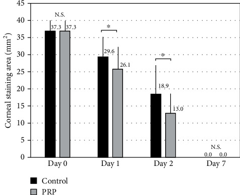
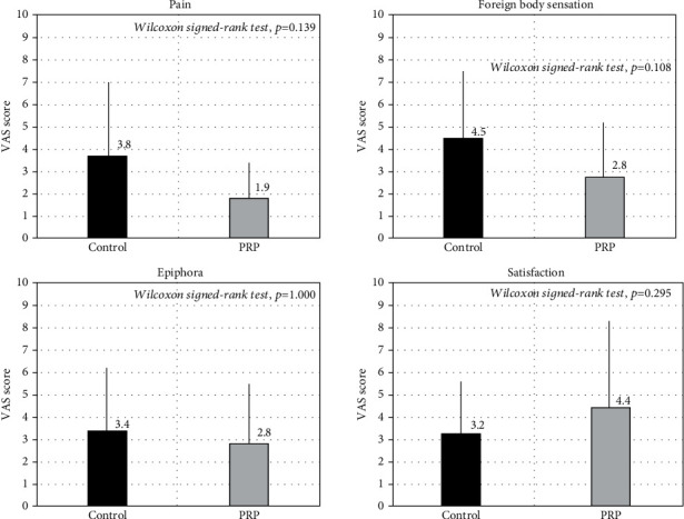
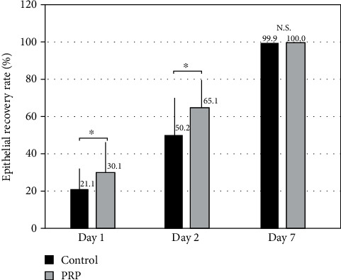
The staining area in the PRP group was significantly smaller than that in the control group on days 1 and 2 (Wilcoxon signed-rank test, = 0.022 and = 0.017, respectively), but not on day 7 ( = 0.317). The recovery rate of the corneal epithelium in the PRP group was significantly higher than that in the control group on days 1 and 2 ( = 0.022 and = 0.017, respectively), but not on day 7 ( = 0.317). We found no significant differences in pain ( = 0.139), foreign body sensation ( = 0.108), epiphora ( = 1.000), or satisfaction ( = 0.295), between the two groups. Postoperative complications did not occur in any of the eyes in the study.
The PRP treatment was effective for enhancing corneal epithelial recovery in the early postoperative period, without significant adverse events, in post-PTK-treated eyes, suggesting that it may hold promise as one of the treatment options for treating such postsurgical patients.
评估富血小板血浆(PRP)对行光动力角膜切削术(PTK)后角膜上皮愈合反应的影响。
我们前瞻性地检查了 10 例患者的 20 只眼,这些患者因颗粒状角膜营养不良或带状角膜病变而行双侧 PTK。患者被随机分配开始每天 4 次局部滴注 PRP 眼用混悬液(PRP 组)或人工泪液(对照组),共 2 周。在 PTK 后即刻、第 1、2 天和第 1 周,我们使用裂隙灯照相术定量测量角膜上皮染色面积。我们还使用视觉模拟评分系统(VAS)来确定主观症状和满意度。
PRP 组在第 1 天和第 2 天的染色面积明显小于对照组(Wilcoxon 符号秩检验, = 0.022 和 = 0.017),但在第 7 天没有差异( = 0.317)。PRP 组角膜上皮的恢复速度明显快于对照组,在第 1 天和第 2 天( = 0.022 和 = 0.017),但在第 7 天没有差异( = 0.317)。我们在两组之间没有发现疼痛( = 0.139)、异物感( = 0.108)、溢泪( = 1.000)或满意度( = 0.295)方面的显著差异。研究中没有任何一只眼发生术后并发症。
PRP 治疗在 PTK 治疗后早期有助于增强角膜上皮的恢复,且无明显不良事件,这表明它可能是治疗此类术后患者的治疗选择之一。