• 文献检索
  • 文档翻译
  • 深度研究
  • 学术资讯
  • Suppr Zotero 插件Zotero 插件
  • 邀请有礼
  • 套餐&价格
  • 历史记录
应用&插件
Suppr Zotero 插件Zotero 插件浏览器插件Mac 客户端Windows 客户端微信小程序
定价
高级版会员购买积分包购买API积分包
服务
文献检索文档翻译深度研究API 文档MCP 服务
关于我们
关于 Suppr公司介绍联系我们用户协议隐私条款
关注我们

Suppr 超能文献

核心技术专利:CN118964589B侵权必究
粤ICP备2023148730 号-1Suppr @ 2026

文献检索

告别复杂PubMed语法,用中文像聊天一样搜索,搜遍4000万医学文献。AI智能推荐,让科研检索更轻松。

立即免费搜索

文件翻译

保留排版,准确专业,支持PDF/Word/PPT等文件格式,支持 12+语言互译。

免费翻译文档

深度研究

AI帮你快速写综述,25分钟生成高质量综述,智能提取关键信息,辅助科研写作。

立即免费体验

抗病毒感知和I型干扰素信号在限制SARS-CoV-2复制中的作用。

Roles of antiviral sensing and type I interferon signaling in the restriction of SARS-CoV-2 replication.

作者信息

Geerling Elizabeth, Pinski Amanda N, Stone Taylor E, DiPaolo Richard J, Zulu Michael Z, Maroney Kevin J, Brien James D, Messaoudi Ilhem, Pinto Amelia K

机构信息

Department of Molecular Microbiology and Immunology, Saint Louis University, St Louis, MO 63103, USA.

Department of Molecular Biology and Biochemistry, University of California-Irvine, Irvine, CA 92697, USA.

出版信息

iScience. 2022 Jan 21;25(1):103553. doi: 10.1016/j.isci.2021.103553. Epub 2021 Dec 3.

DOI:10.1016/j.isci.2021.103553
PMID:34877479
原文链接:https://pmc.ncbi.nlm.nih.gov/articles/PMC8639477/
Abstract

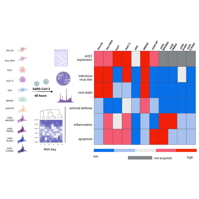
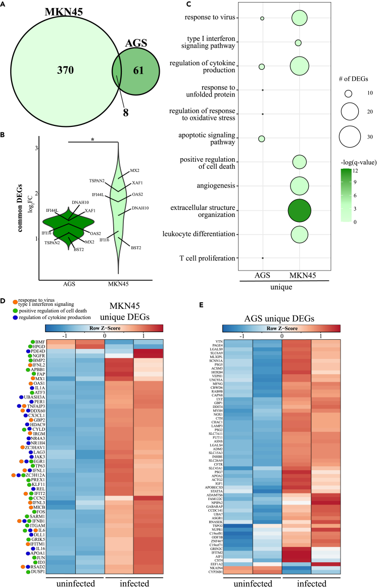
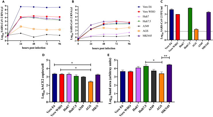
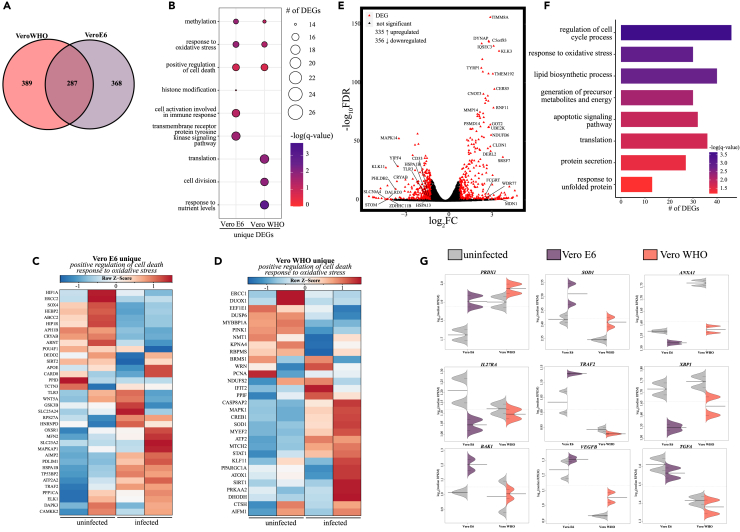
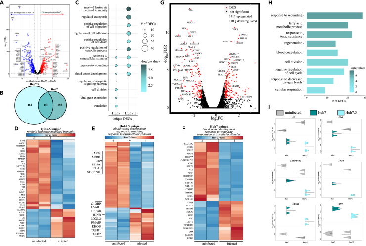
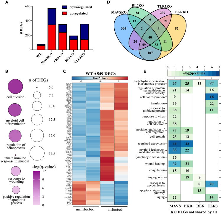
Severe acute respiratory syndrome coronavirus-2 (SARS-CoV-2) is the causative agent of coronavirus disease 2019. Few studies have compared replication dynamics and host responses to SARS-CoV-2 in cell lines from different tissues and species. Therefore, we investigated the role of tissue type and antiviral genes during SARS-CoV-2 infection in nonhuman primate (kidney) and human (liver, respiratory epithelial, gastric) cell lines. We report different viral growth kinetics and release among the cell lines despite comparable ACE2 expression. Transcriptomics revealed that absence of in nonhuman primate cells appeared to enhance inflammatory responses without effecting infectious viral titer. Deletion of in respiratory epithelial cells increased viral replication. Impaired infectious virus release was detected in Huh7 but not Huh7.5 cells, suggesting a role for . Gastric cells MKN45 exhibited robust antiviral gene expression and supported viral replication. Data here provide insight into molecular pathogenesis of and alternative cell lines for studying SARS-CoV-2 infection.

摘要

严重急性呼吸综合征冠状病毒2(SARS-CoV-2)是2019冠状病毒病的病原体。很少有研究比较不同组织和物种的细胞系中SARS-CoV-2的复制动力学和宿主反应。因此,我们研究了组织类型和抗病毒基因在非人类灵长类动物(肾脏)和人类(肝脏、呼吸道上皮、胃)细胞系感染SARS-CoV-2过程中的作用。我们报告了尽管ACE2表达相当,但各细胞系之间的病毒生长动力学和释放情况不同。转录组学显示,非人类灵长类动物细胞中缺乏 似乎增强了炎症反应,而不影响感染性病毒滴度。呼吸道上皮细胞中 的缺失增加了病毒复制。在Huh7细胞中检测到感染性病毒释放受损,但在Huh7.5细胞中未检测到,这表明 起了作用。胃细胞MKN45表现出强大的抗病毒基因表达并支持病毒复制。此处的数据为研究SARS-CoV-2感染的分子发病机制和替代细胞系提供了见解。

https://cdn.ncbi.nlm.nih.gov/pmc/blobs/18e0/8689155/3428baf6e597/gr5.jpg
https://cdn.ncbi.nlm.nih.gov/pmc/blobs/18e0/8689155/15985a77c46d/fx1.jpg
https://cdn.ncbi.nlm.nih.gov/pmc/blobs/18e0/8689155/a01361313d6d/gr1.jpg
https://cdn.ncbi.nlm.nih.gov/pmc/blobs/18e0/8689155/5919ae58e265/gr2.jpg
https://cdn.ncbi.nlm.nih.gov/pmc/blobs/18e0/8689155/a3295c37448d/gr3.jpg
https://cdn.ncbi.nlm.nih.gov/pmc/blobs/18e0/8689155/c68d5521d5f5/gr4.jpg
https://cdn.ncbi.nlm.nih.gov/pmc/blobs/18e0/8689155/3428baf6e597/gr5.jpg

相似文献

1
Roles of antiviral sensing and type I interferon signaling in the restriction of SARS-CoV-2 replication.抗病毒感知和I型干扰素信号在限制SARS-CoV-2复制中的作用。
iScience. 2022 Jan 21;25(1):103553. doi: 10.1016/j.isci.2021.103553. Epub 2021 Dec 3.
2
Antiviral Activity of Type I, II, and III Interferons Counterbalances ACE2 Inducibility and Restricts SARS-CoV-2.I型、II型和III型干扰素的抗病毒活性可抵消ACE2的诱导性并限制新型冠状病毒。
mBio. 2020 Sep 10;11(5):e01928-20. doi: 10.1128/mBio.01928-20.
3
Restriction of SARS-CoV-2 replication by receptor transporter protein 4 (RTP4).通过受体转运蛋白 4(RTP4)限制 SARS-CoV-2 的复制。
mBio. 2023 Aug 31;14(4):e0109023. doi: 10.1128/mbio.01090-23. Epub 2023 Jun 29.
4
Human Nasal and Lung Tissues Infected with SARS-CoV-2 Provide Insights into Differential Tissue-Specific and Virus-Specific Innate Immune Responses in the Upper and Lower Respiratory Tract.人鼻腔和肺部组织感染 SARS-CoV-2 提供了在上呼吸道和下呼吸道中不同组织特异性和病毒特异性先天免疫反应的见解。
J Virol. 2021 Jun 24;95(14):e0013021. doi: 10.1128/JVI.00130-21.
5
Comparative tropism, replication kinetics, and cell damage profiling of SARS-CoV-2 and SARS-CoV with implications for clinical manifestations, transmissibility, and laboratory studies of COVID-19: an observational study.SARS-CoV-2 和 SARS-CoV 的比较嗜性、复制动力学和细胞损伤特征分析及其对 COVID-19 临床表现、传染性和实验室研究的影响:一项观察性研究。
Lancet Microbe. 2020 May;1(1):e14-e23. doi: 10.1016/S2666-5247(20)30004-5. Epub 2020 Apr 21.
6
Angiotensin-Converting Enzyme 2 Potentiates SARS-CoV-2 Infection by Antagonizing Type I Interferon Induction and Its Down-Stream Signaling Pathway.血管紧张素转换酶 2 通过拮抗 I 型干扰素诱导及其下游信号通路增强 SARS-CoV-2 感染。
mSphere. 2022 Aug 31;7(4):e0021122. doi: 10.1128/msphere.00211-22. Epub 2022 Jul 12.
7
SARS-CoV-2 infection and replication kinetics in different human cell types: The role of autophagy, cellular metabolism and ACE2 expression.SARS-CoV-2 感染和复制动力学在不同的人类细胞类型中:自噬、细胞代谢和 ACE2 表达的作用。
Life Sci. 2022 Nov 1;308:120930. doi: 10.1016/j.lfs.2022.120930. Epub 2022 Sep 6.
8
Overcoming Culture Restriction for SARS-CoV-2 in Human Cells Facilitates the Screening of Compounds Inhibiting Viral Replication.克服人细胞中 SARS-CoV-2 的文化限制有助于筛选抑制病毒复制的化合物。
Antimicrob Agents Chemother. 2021 Jun 17;65(7):e0009721. doi: 10.1128/AAC.00097-21.
9
SARS-CoV-2 triggers an MDA-5-dependent interferon response which is unable to control replication in lung epithelial cells.严重急性呼吸综合征冠状病毒2(SARS-CoV-2)引发一种依赖黑色素瘤分化相关基因5(MDA-5)的干扰素反应,这种反应无法控制其在肺上皮细胞中的复制。
J Virol. 2021 Mar 25;95(8). doi: 10.1128/JVI.02415-20. Epub 2021 Jan 29.
10
SARS-CoV-2 Isolates Show Impaired Replication in Human Immune Cells but Differential Ability to Replicate and Induce Innate Immunity in Lung Epithelial Cells.SARS-CoV-2 分离株在人免疫细胞中的复制能力受损,但在肺上皮细胞中的复制能力和诱导先天免疫能力存在差异。
Microbiol Spectr. 2021 Sep 3;9(1):e0077421. doi: 10.1128/Spectrum.00774-21. Epub 2021 Aug 11.

引用本文的文献

1
Influence of Sex and 1,25α Dihydroxyvitamin D on SARS-CoV-2 Infection and Viral Entry.性别与1,25α-二羟维生素D对新型冠状病毒2型感染及病毒进入的影响
Pathogens. 2025 Aug 2;14(8):765. doi: 10.3390/pathogens14080765.
2
Construction and validation of a cell based reporter assay for identifying inhibitors of SARS coronavirus 2 RNA dependent RNA polymerase activity.用于鉴定严重急性呼吸综合征冠状病毒2 RNA依赖性RNA聚合酶活性抑制剂的基于细胞的报告基因检测法的构建与验证
Sci Rep. 2025 May 26;15(1):18443. doi: 10.1038/s41598-025-03813-y.
3
Blood-perfused Vessels-on-Chips stimulated with patient plasma recapitulate endothelial activation and microthrombosis in COVID-19.

本文引用的文献

1
SARS-CoV-2 suppresses IFNβ production mediated by NSP1, 5, 6, 15, ORF6 and ORF7b but does not suppress the effects of added interferon.SARS-CoV-2 抑制由 NSP1、5、6、15、ORF6 和 ORF7b 介导的 IFNβ 产生,但不抑制外加干扰素的作用。
PLoS Pathog. 2021 Aug 26;17(8):e1009800. doi: 10.1371/journal.ppat.1009800. eCollection 2021 Aug.
2
More than 50 long-term effects of COVID-19: a systematic review and meta-analysis.COVID-19 的 50 多种长期影响:系统评价和荟萃分析。
Sci Rep. 2021 Aug 9;11(1):16144. doi: 10.1038/s41598-021-95565-8.
3
Detection of SARS-CoV-2 genome and whole transcriptome sequencing in frontal cortex of COVID-19 patients.
用患者血浆刺激的血液灌注血管芯片可重现新冠病毒疾病中的内皮激活和微血栓形成。
Lab Chip. 2025 Mar 25;25(7):1787-1800. doi: 10.1039/d4lc00848k.
4
Discovering common pathogenetic processes between COVID-19 and tuberculosis by bioinformatics and system biology approach.通过生物信息学和系统生物学方法发现 COVID-19 和结核病之间的共同发病机制。
Front Cell Infect Microbiol. 2023 Dec 15;13:1280223. doi: 10.3389/fcimb.2023.1280223. eCollection 2023.
5
Transcriptional response to VZV infection is modulated by RNA polymerase III in lung epithelial cell lines.病毒 VZV 感染的转录反应受肺上皮细胞系中 RNA 聚合酶 III 的调节。
Front Cell Infect Microbiol. 2022 Jul 25;12:943587. doi: 10.3389/fcimb.2022.943587. eCollection 2022.
6
Plasmalogen Loss in Sepsis and SARS-CoV-2 Infection.脓毒症和新型冠状病毒肺炎感染中的缩醛磷脂损失
Front Cell Dev Biol. 2022 Jun 6;10:912880. doi: 10.3389/fcell.2022.912880. eCollection 2022.
7
Translational Control of COVID-19 and Its Therapeutic Implication.新冠病毒的翻译调控及其治疗意义。
Front Immunol. 2022 Mar 29;13:857490. doi: 10.3389/fimmu.2022.857490. eCollection 2022.
检测 COVID-19 患者额皮质中的 SARS-CoV-2 基因组和全长转录组测序。
Brain Behav Immun. 2021 Oct;97:13-21. doi: 10.1016/j.bbi.2021.05.012. Epub 2021 May 19.
4
Type-I interferon signatures in SARS-CoV-2 infected Huh7 cells.严重急性呼吸综合征冠状病毒2(SARS-CoV-2)感染的Huh7细胞中的I型干扰素特征
Cell Death Discov. 2021 May 18;7(1):114. doi: 10.1038/s41420-021-00487-z.
5
RIG-I triggers a signaling-abortive anti-SARS-CoV-2 defense in human lung cells.RIG-I 在人肺细胞中触发信号转导失败的抗 SARS-CoV-2 防御。
Nat Immunol. 2021 Jul;22(7):820-828. doi: 10.1038/s41590-021-00942-0. Epub 2021 May 11.
6
Overcoming Culture Restriction for SARS-CoV-2 in Human Cells Facilitates the Screening of Compounds Inhibiting Viral Replication.克服人细胞中 SARS-CoV-2 的文化限制有助于筛选抑制病毒复制的化合物。
Antimicrob Agents Chemother. 2021 Jun 17;65(7):e0009721. doi: 10.1128/AAC.00097-21.
7
SARS-CoV-2 induces double-stranded RNA-mediated innate immune responses in respiratory epithelial-derived cells and cardiomyocytes.SARS-CoV-2 诱导呼吸道上皮细胞和心肌细胞中双链 RNA 介导的先天免疫反应。
Proc Natl Acad Sci U S A. 2021 Apr 20;118(16). doi: 10.1073/pnas.2022643118.
8
Multiple sites on SARS-CoV-2 spike protein are susceptible to proteolysis by cathepsins B, K, L, S, and V.SARS-CoV-2 刺突蛋白上的多个位点易受组织蛋白酶 B、K、L、S 和 V 的蛋白水解作用影响。
Protein Sci. 2021 Jun;30(6):1131-1143. doi: 10.1002/pro.4073. Epub 2021 Apr 15.
9
Characterization of SARS-CoV-2 proteins reveals Orf6 pathogenicity, subcellular localization, host interactions and attenuation by Selinexor.严重急性呼吸综合征冠状病毒2(SARS-CoV-2)蛋白的特征揭示了开放阅读框6(Orf6)的致病性、亚细胞定位、宿主相互作用以及塞利尼索对其的减毒作用。
Cell Biosci. 2021 Mar 25;11(1):58. doi: 10.1186/s13578-021-00568-7.
10
SARS-CoV-2 variants reveal features critical for replication in primary human cells.SARS-CoV-2 变体揭示了在原代人细胞中复制的关键特征。
PLoS Biol. 2021 Mar 24;19(3):e3001006. doi: 10.1371/journal.pbio.3001006. eCollection 2021 Mar.