• 文献检索
  • 文档翻译
  • 深度研究
  • 学术资讯
  • Suppr Zotero 插件Zotero 插件
  • 邀请有礼
  • 套餐&价格
  • 历史记录
应用&插件
Suppr Zotero 插件Zotero 插件浏览器插件Mac 客户端Windows 客户端微信小程序
定价
高级版会员购买积分包购买API积分包
服务
文献检索文档翻译深度研究API 文档MCP 服务
关于我们
关于 Suppr公司介绍联系我们用户协议隐私条款
关注我们

Suppr 超能文献

核心技术专利:CN118964589B侵权必究
粤ICP备2023148730 号-1Suppr @ 2026

文献检索

告别复杂PubMed语法,用中文像聊天一样搜索,搜遍4000万医学文献。AI智能推荐,让科研检索更轻松。

立即免费搜索

文件翻译

保留排版,准确专业,支持PDF/Word/PPT等文件格式,支持 12+语言互译。

免费翻译文档

深度研究

AI帮你快速写综述,25分钟生成高质量综述,智能提取关键信息,辅助科研写作。

立即免费体验

小脑卒中后迟发性脑萎缩:地形学关系及临床影响

Delayed cerebral atrophy after cerebellar stroke: topographical relation and clinical impact.

作者信息

van Niftrik Christiaan H B, Visser Thomas F, Sebök Martina, Muscas Giovanni, El Amki Mohamad, Serra Carlo, Regli Luca, Wegener Susanne, Fierstra Jorn

机构信息

Department of Neurosurgery, University Hospital Zurich, University of Zurich, 8091 Zurich, Switzerland.

Clinical Neuroscience Center, University Hospital Zurich, University of Zurich, 8091 Zurich, Switzerland.

出版信息

Brain Commun. 2021 Nov 24;3(4):fcab279. doi: 10.1093/braincomms/fcab279. eCollection 2021.

DOI:10.1093/braincomms/fcab279
PMID:34877537
原文链接:https://pmc.ncbi.nlm.nih.gov/articles/PMC8643502/
Abstract

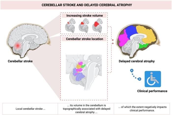
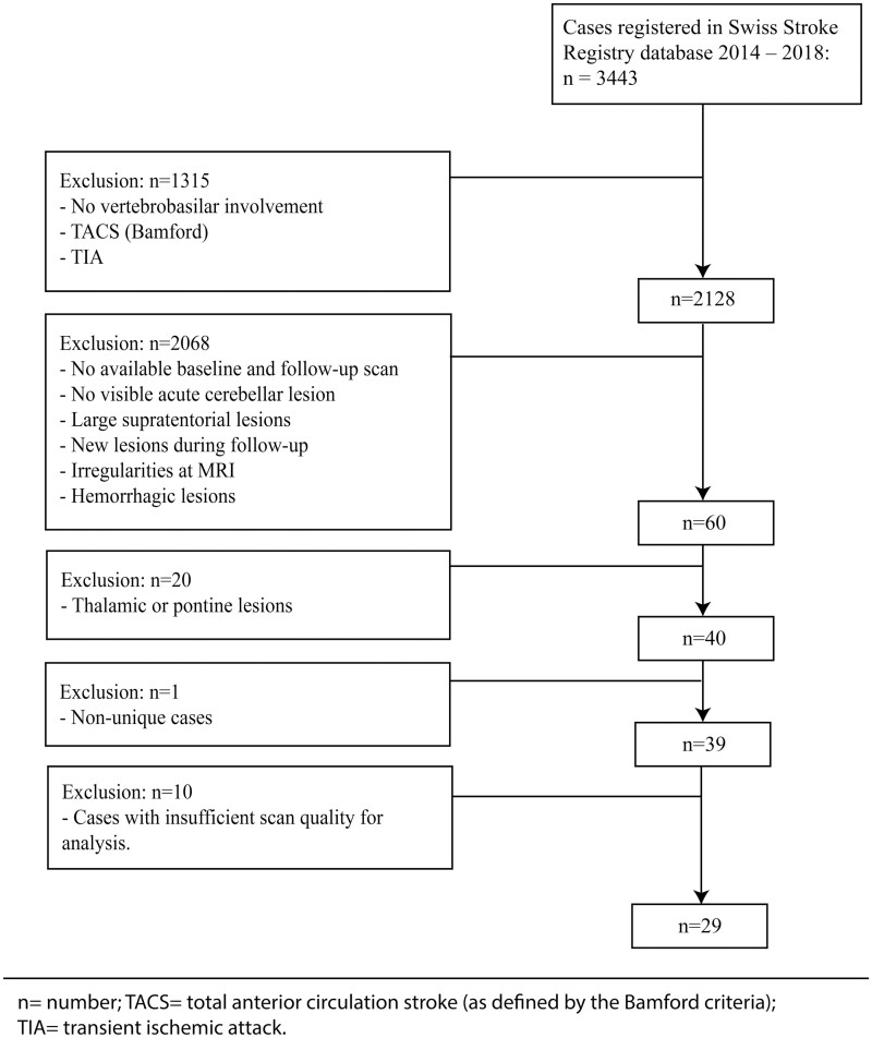
Remote dysconnectivity following cerebellar ischaemic stroke may have a negative impact on supratentorial brain tissue. Since the cerebellum is connected to the individual cerebral lobes via contralateral tracts, cerebellar lesion topography might determine the distribution of contralateral supratentorial brain tissue changes. We investigated (i) the occurrence of delayed cerebral atrophy after cerebellar ischaemic stroke and its relationship to infarct volume; (ii) whether cerebellar stroke topography determines supratentorial atrophy location; and (iii) how cortical atrophy after cerebellar stroke impacts clinical outcome. We performed longitudinal volumetric MRI analysis of patients with isolated cerebellar stroke from the Swiss Stroke Registry database. Stroke location and volume were determined at baseline MRI. Delayed cerebral atrophy was measured as supratentorial cortical volumetric change at follow-up, in contralateral target as compared to ipsilateral reference-areas. In patients with bilateral stroke, both hemispheres were analysed separately. We obtained maps of how cerebellar lesion topography, determines the probability of delayed atrophy per distinct cerebral lobe. Clinical performance was measured with the National Institutes of Health Stroke Scale and modified Rankin Scale. In 29 patients (age 58 ± 18; 9 females; median follow-up: 6.2 months), with 36 datasets (7 patients with bilateral cerebellar stroke), delayed cerebral atrophy occurred in 28 (78%) datasets. A multivariable generalized linear model for a Poisson distribution showed that infarct volume (milliliter) in bilateral stroke patients was positively associated with the number of atrophic target areas (Rate ratio = 1.08; = 0.01). Lobe-specific cerebral atrophy related to distinct topographical cerebellar stroke patterns. By ordinal logistic regression (shift analysis), more atrophic areas predicted higher 3-month mRS scores in patients with low baseline scores (baseline score 3-5: Odds ratio = 1.34; = 0.02; baseline score 0-2: OR = 0.71; = 0.19). Our results indicate that (i) isolated cerebellar ischaemic stroke commonly results in delayed cerebral atrophy and stroke volume determines the severity of cerebral atrophy in patients with bilateral stroke; (ii) cerebellar stroke topography affects the location of delayed cerebral atrophy; and (iii) delayed cerebral atrophy negatively impacts clinical outcome.

摘要

小脑缺血性中风后的远程失连接可能会对幕上脑组织产生负面影响。由于小脑通过对侧传导束与各个脑叶相连,小脑病变的部位可能决定对侧幕上脑组织变化的分布。我们调查了:(i)小脑缺血性中风后迟发性脑萎缩的发生情况及其与梗死体积的关系;(ii)小脑中风部位是否决定幕上萎缩的位置;以及(iii)小脑中风后的皮质萎缩如何影响临床结局。我们对来自瑞士中风登记数据库的孤立性小脑中风患者进行了纵向容积MRI分析。在基线MRI时确定中风部位和体积。迟发性脑萎缩通过随访时幕上皮质容积变化来测量,与同侧参考区域相比,测量对侧目标区域的变化。对于双侧中风患者,分别分析两个半球。我们获得了小脑病变部位如何决定每个不同脑叶迟发性萎缩概率的图谱。临床表现在国立卫生研究院卒中量表和改良Rankin量表上进行测量。在29例患者(年龄58±18岁;9名女性;中位随访时间:6.2个月)中,有36个数据集(7例双侧小脑中风患者),28个(78%)数据集出现了迟发性脑萎缩。一个用于泊松分布的多变量广义线性模型显示,双侧中风患者的梗死体积(毫升)与萎缩目标区域的数量呈正相关(率比=1.08; =0.01)。特定脑叶的脑萎缩与不同的小脑中风部位模式相关。通过有序逻辑回归(移位分析),在基线评分较低的患者中,更多的萎缩区域预测3个月时改良Rankin量表评分更高(基线评分3 - 5分:优势比=1.34; =0.02;基线评分0 - 2分:优势比=0.71; =0.19)。我们的结果表明:(i)孤立性小脑缺血性中风通常会导致迟发性脑萎缩,中风体积决定了双侧中风患者脑萎缩的严重程度;(ii)小脑中风部位影响迟发性脑萎缩的位置;以及(iii)迟发性脑萎缩对临床结局有负面影响。

https://cdn.ncbi.nlm.nih.gov/pmc/blobs/d489/8643502/6b996294908e/fcab279f3.jpg
https://cdn.ncbi.nlm.nih.gov/pmc/blobs/d489/8643502/4b6b4e4a4f9c/fcab279f4.jpg
https://cdn.ncbi.nlm.nih.gov/pmc/blobs/d489/8643502/4b40d1fec519/fcab279f1.jpg
https://cdn.ncbi.nlm.nih.gov/pmc/blobs/d489/8643502/380becb19f39/fcab279f2.jpg
https://cdn.ncbi.nlm.nih.gov/pmc/blobs/d489/8643502/6b996294908e/fcab279f3.jpg

相似文献

1
Delayed cerebral atrophy after cerebellar stroke: topographical relation and clinical impact.小脑卒中后迟发性脑萎缩:地形学关系及临床影响
Brain Commun. 2021 Nov 24;3(4):fcab279. doi: 10.1093/braincomms/fcab279. eCollection 2021.
2
Crossed cerebellar hypoperfusion in hyperacute ischemic stroke.超急性缺血性卒中的交叉性小脑灌注不足
J Neurol Sci. 2004 Oct 15;225(1-2):65-9. doi: 10.1016/j.jns.2004.07.004.
3
Quantitative longitudinal evaluation of diaschisis-related cerebellar perfusion and diffusion parameters in patients with supratentorial hemispheric high-grade gliomas after surgery.幕上半球高级别胶质瘤患者术后与交叉性小脑失联络相关的小脑灌注及扩散参数的定量纵向评估
Cerebellum. 2014 Oct;13(5):580-7. doi: 10.1007/s12311-014-0575-2.
4
Crossed cerebellar diaschisis and brain recovery after stroke.交叉性小脑去传入和中风后的脑恢复
Stroke. 1995 Jan;26(1):90-5. doi: 10.1161/01.str.26.1.90.
5
Detection of Crossed Cerebellar Diaschisis by Intravoxel Incoherent Motion MR Imaging in Subacute Ischemic Stroke.脑桥交叉性小脑失联络的磁共振体素内不相干运动弥散加权成像研究。
Cell Transplant. 2019 Aug;28(8):1062-1070. doi: 10.1177/0963689719856290. Epub 2019 Jun 14.
6
Crossed cerebellar diaschisis and crossed cerebellar atrophy: correlation of MR findings, clinical symptoms, and supratentorial diseases in 26 patients.交叉性小脑神经机能联系失联络和交叉性小脑萎缩:26例患者的磁共振成像表现、临床症状与幕上疾病的相关性
AJR Am J Roentgenol. 1992 May;158(5):1155-9. doi: 10.2214/ajr.158.5.1566683.
7
Crossed cerebellar diaschisis in patients with acute middle cerebral artery infarction: Occurrence and perfusion characteristics.急性大脑中动脉梗死患者的交叉性小脑失联络:发生率及灌注特征
J Cereb Blood Flow Metab. 2016 Apr;36(4):743-54. doi: 10.1177/0271678X15617953. Epub 2015 Dec 1.
8
Remote regional cerebral blood flow consequences of focused infarcts of the medulla, pons and cerebellum.延髓、脑桥和小脑局灶性梗死对远隔区域脑血流的影响
J Nucl Med. 1999 May;40(5):721-9.
9
Characteristics of cerebral perfusion and diffusion associated with crossed cerebellar diaschisis after acute ischemic stroke.急性缺血性卒中后交叉性小脑性失联络相关的脑灌注和扩散特征
Jpn J Radiol. 2020 Feb;38(2):126-134. doi: 10.1007/s11604-019-00898-0. Epub 2019 Nov 13.
10
Brain Atrophy and the Risk of Futile Endovascular Reperfusion in Acute Ischemic Stroke.脑萎缩与急性缺血性脑卒中血管内再通治疗无效的风险。
Stroke. 2020 May;51(5):1514-1521. doi: 10.1161/STROKEAHA.119.028511. Epub 2020 Mar 19.

引用本文的文献

1
Impaired dynamic cerebral autoregulation measured in the middle cerebral artery in patients with vertebrobasilar ischemia is associated with autonomic failure.在椎基底动脉缺血患者的大脑中动脉中测量到的动态脑自动调节功能障碍与自主神经衰竭有关。
J Stroke Cerebrovasc Dis. 2024 Jan;33(1):107454. doi: 10.1016/j.jstrokecerebrovasdis.2023.107454. Epub 2023 Nov 4.
2
Cognitive impairment in essential tremor assessed by the cerebellar cognitive affective syndrome scale.通过小脑认知情感综合征量表评估特发性震颤中的认知障碍。
Front Neurol. 2023 Aug 18;14:1224478. doi: 10.3389/fneur.2023.1224478. eCollection 2023.
3
Determinant of the cerebellar cognitive affective syndrome in Friedreich's ataxia.

本文引用的文献

1
Crossed Cerebellar Diaschisis in Patients With Symptomatic Unilateral Anterior Circulation Stroke Is Associated With Hemodynamic Impairment in the Ipsilateral MCA Territory.有症状的单侧前循环卒中患者的交叉性小脑失联络与同侧大脑中动脉区域的血流动力学损害有关。
J Magn Reson Imaging. 2021 Apr;53(4):1190-1197. doi: 10.1002/jmri.27410. Epub 2020 Oct 28.
2
Crossed Cerebellar Diaschisis Indicates Hemodynamic Compromise in Ischemic Stroke Patients.交叉性小脑 diaschisis 提示缺血性脑卒中患者存在血液动力学障碍。
Transl Stroke Res. 2021 Feb;12(1):39-48. doi: 10.1007/s12975-020-00821-0. Epub 2020 Jun 6.
3
Whole-body somatotopic maps in the cerebellum revealed with 7T fMRI.
弗里德赖希共济失调中小脑认知情感综合征的决定因素。
J Neurol. 2023 Jun;270(6):2969-2974. doi: 10.1007/s00415-023-11623-3. Epub 2023 Feb 15.
4
Cerebellar cognitive affective syndrome after acute cerebellar stroke.急性小脑卒中后的小脑认知情感综合征
Front Neurol. 2022 Aug 11;13:906293. doi: 10.3389/fneur.2022.906293. eCollection 2022.
5
Fully Automatic Classification of Brain Atrophy on NCCT Images in Cerebral Small Vessel Disease: A Pilot Study Using Deep Learning Models.脑小血管病中非增强CT图像上脑萎缩的全自动分类:一项使用深度学习模型的初步研究
Front Neurol. 2022 Mar 24;13:846348. doi: 10.3389/fneur.2022.846348. eCollection 2022.
7T fMRI 揭示小脑的全身躯体感觉图谱。
Neuroimage. 2020 May 1;211:116624. doi: 10.1016/j.neuroimage.2020.116624. Epub 2020 Feb 11.
4
Functional tracts of the cerebellum-essentials for the neurosurgeon.小脑的功能束——神经外科医生的必备知识
Neurosurg Rev. 2021 Feb;44(1):273-278. doi: 10.1007/s10143-020-01242-1. Epub 2020 Feb 13.
5
Functional boundaries in the human cerebellum revealed by a multi-domain task battery.人类小脑的功能边界由多领域任务电池揭示。
Nat Neurosci. 2019 Aug;22(8):1371-1378. doi: 10.1038/s41593-019-0436-x. Epub 2019 Jul 8.
6
Characterizing ipsilateral thalamic diaschisis in symptomatic cerebrovascular steno-occlusive patients.描述症状性脑血管狭窄闭塞患者的同侧丘脑失联络现象。
J Cereb Blood Flow Metab. 2020 Mar;40(3):563-573. doi: 10.1177/0271678X19830532. Epub 2019 Feb 12.
7
NIHSS cut point for predicting outcome in supra- vs infratentorial acute ischemic stroke.NIHSS 截断值预测幕上与幕下急性缺血性脑卒中的结局。
Neurology. 2018 Oct 30;91(18):e1695-e1701. doi: 10.1212/WNL.0000000000006437. Epub 2018 Sep 28.
8
BOLD cerebrovascular reactivity as a novel marker for crossed cerebellar diaschisis.脑血容量反应性作为交叉性小脑失联络的新型标志物。
Neurology. 2018 Oct 2;91(14):e1328-e1337. doi: 10.1212/WNL.0000000000006287. Epub 2018 Sep 5.
9
Differences in motor network dynamics during recovery between supra- and infra-tentorial ischemic strokes.幕上和幕下缺血性卒中后恢复期间运动网络动力学的差异。
Hum Brain Mapp. 2018 Dec;39(12):4976-4986. doi: 10.1002/hbm.24338. Epub 2018 Aug 18.
10
Functional gradients of the cerebellum.小脑的功能梯度。
Elife. 2018 Aug 14;7:e36652. doi: 10.7554/eLife.36652.