Tehran University of Medical Sciences, School of Dentistry, Oral and Maxillofacial Pathology Department, Tehran, Iran.
Tehran University of Medical Sciences, School of Dentistry, Tehran, Iran.
J Appl Oral Sci. 2021 Dec 1;29:e20210374. doi: 10.1590/1678-7757-2021-0374. eCollection 2021.
Squamous cell carcinoma antigen (SCCA) is used as a prognostic marker for recurrence of squamous cell carcinoma in various sites, including head and neck. Studies suggest that its high serum levels are correlated to some clinical features, such as nodal metastasis. However, it is still unknown if high SCCA in patients with SCCA tissue expression in tumor cells are related to peripheral T-lymphocytes. Therefore, we did this study to evaluate SCCA expression in squamous cell carcinoma and verrucous carcinoma and to compare it with normal oral mucosa, also investigating the correlation between serum-based and tissue-based antigen levels.
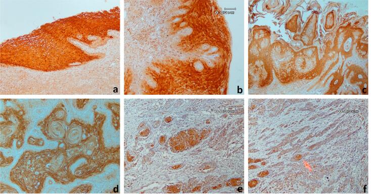
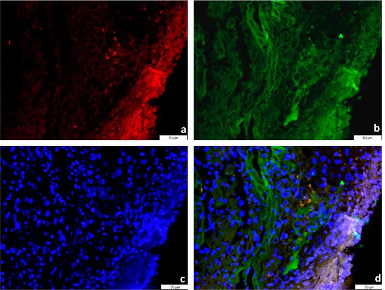
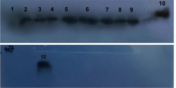
In this study, the immunohistochemistry (IHC) technique was used to determine the SCCA1 expression pattern in 81 specimens divided into 3 groups, including oral squamous cell carcinoma, verrucous carcinoma, and normal oral mucosa. Serum-based and tissue-based antigen levels of 20 oral squamous cell carcinoma cases were compared by the western blot assay. SCCA expression was also evaluated and compared in both tumor cells and peripheral T-lymphocytes by the immunofluorescence assay.
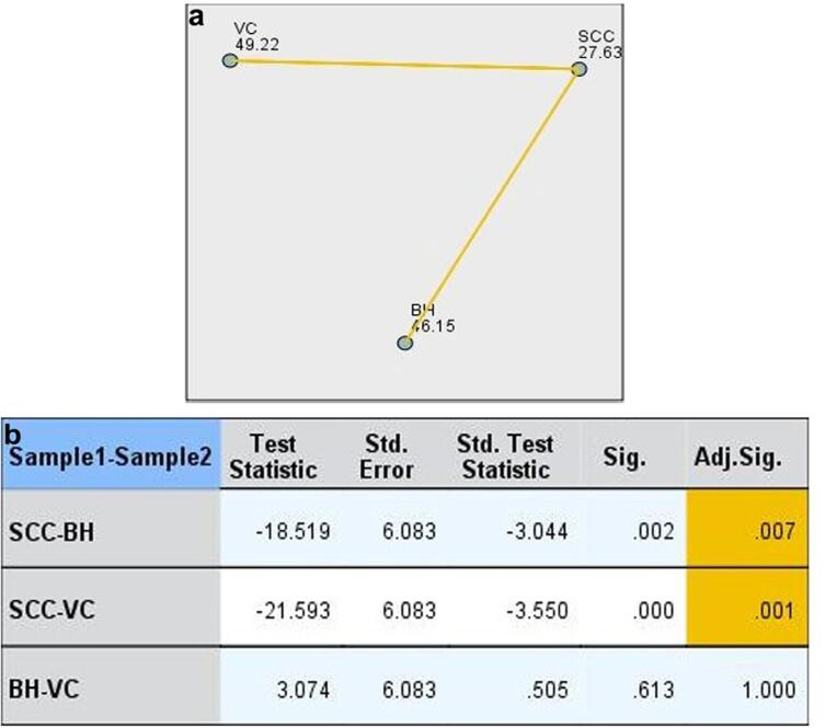
Our results showed that the SCCA levels in SCC specimens were significantly lower than in verrucous carcinoma and normal and hyperplastic oral mucosa specimens. We found no correlation between the IHC expression of SCCA and serum levels. SCCA was well expressed in both tumor cells and peripheral T-lymphocytes.
Decreasing SCCA in SCC specimens suggested that SCC tumor cells may affect more than the serum levels of SCCA in some patients. In addition, expression of SCCA in peripheral T-lymphocytes showed that both tumor cells and T-lymphocytes may cause serum SCCA.
鳞状细胞癌抗原(SCCA)被用作各种部位鳞状细胞癌(包括头颈部)复发的预后标志物。研究表明,其血清水平高与一些临床特征相关,如淋巴结转移。然而,SCCA 组织表达高的患者的 SCCA 与外周 T 淋巴细胞之间的关系仍不清楚。因此,我们进行了这项研究,以评估 SCCA 在鳞状细胞癌和疣状癌中的表达,并将其与正常口腔黏膜进行比较,同时还研究了基于血清和基于组织的抗原水平之间的相关性。
在这项研究中,我们使用免疫组织化学(IHC)技术来确定 81 个标本中的 SCCA1 表达模式,这些标本分为 3 组,包括口腔鳞状细胞癌、疣状癌和正常口腔黏膜。通过 Western blot 检测比较了 20 例口腔鳞状细胞癌病例的基于血清和基于组织的抗原水平。我们还通过免疫荧光法评估并比较了 SCCA 在肿瘤细胞和外周 T 淋巴细胞中的表达。
我们的结果表明,SCCA 水平在 SCC 标本中明显低于疣状癌和正常及增生性口腔黏膜标本。我们没有发现 SCCA 的 IHC 表达与血清水平之间存在相关性。SCCA 在肿瘤细胞和外周 T 淋巴细胞中均有良好表达。
SCCA 水平在 SCC 标本中降低表明,SCCA 肿瘤细胞可能影响一些患者的血清 SCCA 水平。此外,外周 T 淋巴细胞中 SCCA 的表达表明肿瘤细胞和 T 淋巴细胞均可能导致血清 SCCA。